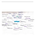
4BBY1020- Chemistry for the Biosciences
L1: Introductions to solutions chemistry and calculations
• mass = conc x vol • 1mg/mL = 1g/L
• mass = mol = mr • 1mg/L = 0.001g/L
• g/L = M x mr • 1µg/mL = 0.001mg/mL = 0.001g/L
• % (w/v) = g/100mL and % (v/v) = mL/100mL. e.g 60g/L = 6g/100mL = 6% (w/v).
• C1V1 = C2V2 is used for all dilutions. e.g 1 in 104 dilution means V2 = 104V.
L2: Atoms, compounds and chemical bonding
• When an electron jumps to a lower shell a photon is emitted, the higher the shell it goes to the
higher energy the photon has.
• Electrons are added to orbitals based on the Aufbau principle,
Pauli exclusion and Hund’s rule.
• 4 different types of orbitals, s, p, d and f. These can then be further
split into other types depending on their orientations against a
specific axis. 4s2 comes before 3d10.
• e.g N = 1s2 2s2 2p3
• The energy within each shell = principal quantum number (n) + orbital quantum number (l).
• Valency = number of bonds + formal charge. The highest occupied shell is the valence shell for
that atom. The number of free spaces within that shell is its valency. e.g for N valency = 3.
• Electronegativity (Δx) is a chemical property that describes the ability of
an atom to attract electrons towards itself. If Δx >1.7 = ionic bonding
and if Δx < 0.7 = covalent bonding.
• Covalent bonding: Two atoms come together and merge to form new
molecular orbitals. One forms a low energy bonding orbital and one
forms a high energy anti-bonding orbital. If there are electrons in the
anti-bonding orbital covalent bonds will not form. Easy to see with
Lewis structures.
• Lone pairs occupy non-bonding molecular orbitals. A dative covalent
bond is where both electrons within the covalent bond come from the
same atom.
• Two s orbitals will form a σ bond. Two p orbitals will form a π bond.
• π bonds help form conjugated systems, a system of atoms covalently bonded with alternating
single and multiple bonds in a molecule of an organic compound, resulting in delocalisation/
resonance, where a compound can be represented with more than one Lewis structure.
• Aromatic compounds consist of a conjugated ring of C-C bonds with resonance due to p
orbitals on adjacent carbon atoms overlapping. Any planar ring with (4n+2)π electrons is
aromatic e.g paracetamol, ibuprofen, tyrosine, phenylalanine, benzene etc.
L3: Molecular shape and forces: non-covalent interactions
• Molecular geometry is determined by bond length, bond angles and bond rotation.
• The bond lengths between atoms are normally based on the atomic radii of the atoms.
• Single bonds are longer and weaker than double or triple bonds.
• VESPR theory suggests that pairs of valence electrons repel each other, influencing geometry.
, • Hybridisation (4 domains = sp3 etc) where 5 = sp3d and 6 = sp3d2 including lone pairs.
• Dihedral angles ɸ (phi) and Ѱ (psi) describe the rotation about the N-C and C-C bonds in amino
acids. These help contribute to the twisting backbone of secondary structure.
• Molecular dipoles are formed when two atoms with a high difference in electronegativity form a
covalent bond. e.g in HF, an electron cloud will be partly negative at F and partly positive at H.
• Water molecules have a permanent dipole meaning it is a polar solvent. Even if bonds are polar
doesn’t mean the whole molecule is.
• London Dispersion forces happen between all molecules, causing an instantaneous dipole.
• Hydrogen bonds occur when an H is covalently bonded to an F, O or N.
• Ion-dipole interactions allow the solubility of NaCl in water.
L4: pH and buffering
• pH is a measure of [H+]. The acidity of a solution depends only on free hydrogen ions.
• Regulation of blood pH is critical (7.35-7.45). The living range is 7.0-7.8 otherwise acidosis or
alkalosis occurs.
• Most H+ is generated from breakdown of proteins, incomplete oxidation of fats or glucose and
the loading and transport of CO2 in the blood.
• Acid-base balance is regulated in the body by the lungs, kidneys and chemical buffers in the
blood.
• Buffers resist abrupt and large swings in the pH of body fluids by releasing H+ when pH begins
to rise and binding H+ when the pH drops.
pH = -log [H+], meaning when [H+] is 10-2 the pH is 2.
• At neutrality, [H ] = [OH-] so [H+] is 10-7 and pH is 7 as the ionic product of water is 10-4 M2.
+
• Blood pH is 7.4, meaning the [H+] is 3.98 x 10⁻⁸ M.
• Acids are proton donors and bases are proton acceptors.
• Acids that dissociate completely in solution are strong acids (HCl→H+ + Cl-) whereas those that
dissociate incompletely are weak acids (H2CO3→ H+ + HCO3-).
• Increasing the pH of a solution (e.g adding NaOH) then more of the weak acid will dissociate.
• The pH at which weak acid is half dissociated is known as the pKa. The weaker the acid, the
higher the pKa.
pKa = -log Ka, where Ka is the dissociation constant.
[A −] [con jugateba se]
pH = pKa + log = pKa + log
[H A] [acid ]
• Buffers are mixtures of weak acids and their conjugate bases. At pKa, buffering is optimal.
• Physiologically important buffers must be able to dissociate at physiological pH.
• In blood, saliva, other body fluids H2CO3→H+ + HCO3- (pKa 6.1) and H2PO4-→HPO42- (pKa 6.8).
• Proteins can also be used as buffers. e.g histidine has a pKa of 6.
• H2CO3 is proportional to the PCO2.
• Haemoglobin is a good buffer as it has a large number of histidine residues. The surroundings of
an acid group influence the pKa. Whilst free histidine has a pKa of 6, in oxyhemoglobin pKa is
6.8 and in deoxyhemoglobin pKa is 7.8.
L5: Carbon compounds and isomerisation
• Polarity of functional groups (more polar = higher boiling point):
amide > acid > alcohol > ketone ~ aldehyde > amide > ester > ether > alkane
-meth -eth -prop -but -pent -hex -hept -oct -non -dec