Case Study of Patient with ACS_CAD_CABG.
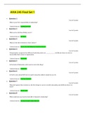
Case Study of Patient with ACS_CAD_CABG. Scenario Your patient, 58-year-old K.Z., has a significant cardiac history. He has long-standing coronary artery dis- ease (CAD) with occasional episodes of heart failure (HF). One year ago, he had an anterior wall myocar- dial infarction (MI). In addition, he has chronic anemia, hypertension, chronic renal insufficiency, and a recently diagnosed 4-cm suprarenal abdominal aortic aneurysm. Because of his severe CAD, he had to retire from his job as a railroad engineer about 6 months ago. This morning, he is being admitted to your telemetry unit for a same-day cardiac catheterization. As you take his health history, you note that his wife died a year ago (at about the same time that he had his MI) and he does not have any children. He is a current cigarette smoker with a 50–pack-year smoking history. His vital signs (VS) are 158/94, 88, 20, and 97.2 ° F (36.2 ° C). As you talk with him, you realize that he has only a minimal understanding of the catheterization procedure. 1. Before he leaves for the catheterization laboratory, you briefly teach him the important things he needs to know before having the procedure. List five priority topics you will address. 2. Look at his past history. What other factors are present that could contribute to his risk for cardiac ischemia? CASE STUDY PROGRESS Several hours later, K.Z. returns from his catheterization. The catheterization report shows 90% occlusion of the proximal left anterior descending (LAD) coronary artery, 90% occlusion of the distal LAD, 70% to 80% occlusion of the distal right coronary artery (RCA), an old apical infarct, and an ejection fraction (EF) of 37%. About an hour after the procedure is finished, you perform a brief physical assessment and note a grade III/VI systolic ejection murmur at the cardiac apex, crackles bilaterally in the lung bases, and trace itting edema of his feet and ankles. Except for the soft systolic murmur, these findings were not present before the catheterization. 3. Using the following diagram, identify the superior vena cava, the aorta, and the left and right ventricles. Identify the main coronary arteries, and circle the areas of the LAD and RCA that have significant occlusion, as identified in the previous report. Lightly shade the area of the heart where K.Z. had the earlier infarct.
Written for
- Institution
-
South Texas College
- Module
-
RNSG 1417
Document information
- Uploaded on
- January 10, 2022
- Number of pages
- 3
- Written in
- 2021/2022
- Type
- Case
- Professor(s)
- Professor
- Grade
- A+
Subjects
-
case study of patient with acscadcabg scenario your patient
-
58 year old kz
-
has a significant cardiac history he has long standing coronary artery dis ease cad with occasional episodes of h