Cells and Microscopes
Prokaryotic vs Eukaryotic cells
- Eukaryotic cells have a membrane- bound nucleus and
prokaryotic do not
- Prokaryotic cells have no membrane - bound structures, they
tend to be small, simple cells, measuring around 0.1 - 5µm in
diameter
- While prokaryotic cells do not have membrane-bound
structures, they do have distinct cellular regions. In prokaryotic
cells, DNA bundles together in a region called the nucleoid.
- eg. bacteria and archaea
- eukaryotes are organisms whose cells have a nucleus and
other organelles enclosed by a plasma membrane.
- Eukaryotic cells are large (around 10-100 μm) and complex.
While most eukaryotes are multicellular organisms, there are some single-cell eukaryotes.
- eg. animals, plants, fungi, algae and protozoans
- info: technology networks : cell science
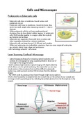
Laser Scanning Confocal Microscopy
- optical imaging technique for increasing optical resolution and
contrast of a micrograph by using a spatial pinhole to block out of
focus light in the image formation
- CLSM combines high-resolution optical imaging with depth selectivity
which allows us to do optical sectioning. This means that we can view
visual sections of tiny structures that would be difficult to physically
section (e.g. embryos) and construct 3-D structures from the obtained
images.
- The CLSM works by passing a laser beam through a light source aperture
which is then focused by an objective lens into a small area on the surface of your sample and
an image is built up pixel-by-pixel by collecting the emitted photons from the fluorophores in the
sample.
Prokaryotic vs Eukaryotic cells
- Eukaryotic cells have a membrane- bound nucleus and
prokaryotic do not
- Prokaryotic cells have no membrane - bound structures, they
tend to be small, simple cells, measuring around 0.1 - 5µm in
diameter
- While prokaryotic cells do not have membrane-bound
structures, they do have distinct cellular regions. In prokaryotic
cells, DNA bundles together in a region called the nucleoid.
- eg. bacteria and archaea
- eukaryotes are organisms whose cells have a nucleus and
other organelles enclosed by a plasma membrane.
- Eukaryotic cells are large (around 10-100 μm) and complex.
While most eukaryotes are multicellular organisms, there are some single-cell eukaryotes.
- eg. animals, plants, fungi, algae and protozoans
- info: technology networks : cell science
Laser Scanning Confocal Microscopy
- optical imaging technique for increasing optical resolution and
contrast of a micrograph by using a spatial pinhole to block out of
focus light in the image formation
- CLSM combines high-resolution optical imaging with depth selectivity
which allows us to do optical sectioning. This means that we can view
visual sections of tiny structures that would be difficult to physically
section (e.g. embryos) and construct 3-D structures from the obtained
images.
- The CLSM works by passing a laser beam through a light source aperture
which is then focused by an objective lens into a small area on the surface of your sample and
an image is built up pixel-by-pixel by collecting the emitted photons from the fluorophores in the
sample.