4.3 Protein Tertiary and Quaternary Structures
SUMMARY 4.3 Protein Tertiary and Quaternary Structures
■ Tertiary structure is the complete three-dimensional structure of a polypeptide chain.
Many proteins fall into one of two general classes of proteins based on tertiary
structure: fibrous and globular.
■ Fibrous proteins, which serve mainly structural roles, have simple repeating elements
of secondary structure.
■ Globular proteins have more complicated tertiary structures, often containing several
types of secondary structure in the same polypeptide chain. The first globular protein
structure to be determined, by x-ray diffraction methods, was that of myoglobin.
■ The complex structures of globular proteins can be analyzed by examining folding
patterns called motifs (also called folds or supersecondary structures). The many
thousands of known protein structures are generally assembled from a repertoire of
only a few hundred motifs. Domains are regions of a polypeptide chain that can fold
stably and independently.
■ Some proteins or protein segments are intrinsically disordered, lacking definable
three-dimensional structure. These proteins have distinctive amino acid compositions
that allow a more flexible structure. Some of these disordered proteins function as
structural components or scavengers; others can interact with many different protein
partners, serving as versatile inhibitors or as central components of protein interaction
networks. Quaternary structure results from interactions between the subunits of
multisubunit (multimeric) proteins or large protein assemblies. Some multimeric
proteins have a repeated unit consisting of a single subunit or a group of subunits, each
unit called a protomer.
The overall three-dimensional arrangement of all atoms in a protein is referred to as the
protein’s tertiary structure. Whereas the term “secondary structure” refers to the
spatial arrangement of amino acid residues that are adjacent in a segment of a
polypeptide, tertiary structure includes longer- range aspects of amino acid sequence.
Amino acids that are far apart in the polypeptide sequence and are in different types of
secondary structure may interact within the completely folded structure of a protein.
The location of bends (including β turns) in the polypeptide chain and the direction and
angle of these bends are determined by the number and location of specific bend-
producing residues, such as Pro, Thr, Ser, and Gly. Interacting segments of polypeptide
chains are held in their characteristic tertiary positions by several kinds of weak
interactions (and sometimes by covalent bonds such as disulfide cross-links) between
the segments.
Some proteins contain two or more separate polypeptide chains, or subunits, which may
be identical or different. The arrangement of these protein subunits in three-
dimensional complexes constitutes quaternary structure.
,In considering these higher levels of structure, it is useful to designate two major groups
into which many proteins can be classified: fibrous proteins, with polypeptide chains
arranged in long strands or sheets, and globular proteins, with polypeptide chains
folded into a spherical or globular shape. The two groups are structurally distinct.
Fibrous proteins usually consist largely of a single type of secondary structure, and their
tertiary structure is relatively simple. Globular proteins often contain several types of
secondary structure. The two groups also differ functionally: the structures that provide
support, shape, and external protection to vertebrates are made of fibrous proteins,
whereas most enzymes and regulatory proteins are globular proteins.
The oxygen nucleus attracts electrons more strongly than does the hydrogen nucleus (a
proton); that is, oxygen is more electronegative. The nearly tetrahedral arrangement of
the orbitals about the oxygen atom (Fig. 2-1a) allows each water molecule to form hydrogen
bonds with as many as four neighboring water molecules. Uncharged but polar biomolecules
such as sugars dissolve readily in water because of the stabilizing effect of hydrogen bonds
between the hydroxyl groups or carbonyl oxygen of the sugar and the polar water molecules
Fibrous Proteins Are Adapted for a Structural Function
α-Keratin, collagen, and silk fibroin nicely illustrate the relationship between protein
structure and biological function (Table 4-3). Fibrous proteins share properties that give
strength and/or flexibility to the structures in which they occur. In each case, the
fundamental structural unit is a simple repeating
element of secondary structure. All fibrous proteins are insoluble in water, a property
conferred by a high concentration of hydrophobic amino acid residues both in the
interior of the protein and on its surface. These hydrophobic surfaces are largely buried,
as many similar polypeptide chains are packed together to form elaborate
supramolecular complexes. The underlying structural simplicity of fibrous proteins
makes them particularly useful for illustrating some of the fundamental principles of
protein structure discussed above.
4-3 Secondary Structures and Properties of Some Fibrous Proteins
α-Keratin The α-keratins have evolved for strength. Found only in mammals, these
proteins constitute almost the entire dry weight of hair, wool, nails, claws, quills, horns,
hooves, and much of the outer layer of skin. The α- keratins are part of a broader family
of proteins called intermediate filament (IF) proteins. Other IF proteins are found in the
cytoskeletons of animal cells. All IF proteins have a structural function and share the
structural features exemplified by the α-keratins.
The α-keratin helix is a right-handed α helix, the same helix found in many other
proteins. Francis Crick and Linus Pauling, in the early 1950s, independently suggested
that the α helices of keratin were arranged as a coiled coil. Two strands of α-keratin,
oriented in parallel (with their amino termini at the same end), are wrapped about each
other to form a supertwisted
Collagen triple High tensile strength, without Collagen of helix stretch tendons, bone
matrix coiled coil. The supertwisting amplifies the strength of the overall structure, just
as strands are twisted to make a strong rope (Fig. 4-11). The twisting of the axis of an α
helix to form a coiled coil explains the discrepancy between the 5.4 Å per turn predicted
, for an α helix by Pauling and Corey and the 5.15 to 5.2 Å repeating structure observed
in the x-ray diffraction of hair (p. 152). The helical path of the supertwists is left-handed,
opposite in sense to the α helix. The surfaces where the two α helices touch are made
up of hydrophobic amino acid residues, their R groups meshed together in a regular
interlocking pattern. This permits a close packing of the polypeptide chains within the
left-handed supertwist. Not surprisingly, α-keratin is rich in the hydrophobic residues
Ala, Val, Leu, Ile, Met, and Phe.
Water is effective in screening the electrostatic interactions between dissolved ions because
it has a high dielectric constant, a physical property
An individual polypeptide in the α-keratin coiled coil has a relatively simple tertiary
structure, dominated by an α-helical secondary structure with its helical axis twisted in
a left-handed superhelix. The intertwining of the two α-helical polypeptides is an
example of quaternary structure. Coiled coils of this type are common structural
elements in filamentous proteins and in the muscle protein myosin (see Fig. 5-27). The
quaternary structure of α- keratin can be quite complex. Many coiled coils can be
assembled into large supramolecular complexes, such as the arrangement of α-keratin
that forms the intermediate filament of hair (Fig. 4-11b).
The strength of fibrous proteins is enhanced by covalent cross-links between
polypeptide chains in the multihelical “ropes” and between adjacent chains in a
supramolecular assembly. In α-keratins, the cross-links stabilizing quaternary structure
are disulfide bonds (Box 4-2). In the hardest and toughest α-keratins, such as those of
rhinoceros horn, up to 18% of the residues are cysteines involved in disulfide bonds.
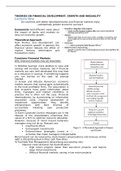
FIGURE 4-11 Structure of hair. (a) Hair α-keratin is an elongated α helix with somewhat
thicker elements near the amino and carboxyl termini. Pairs of these helices are
interwound in a left-handed sense to form two-chain coiled coils. These then combine in
higher-order structures called protofilaments and protofibrils. About four protofibrils—32
strands of α-keratin in all—combine to form an intermediate filament. The individual
two-chain coiled coils in the various substructures also seem to be interwound, but the
handedness of the interwinding and other structural details are unknown. (b) A hair is
an array of many α-keratin filaments, made up of the substructures shown in (a)
Box 4.2 – Permanent Waving Is Biochemical
Engineering When hair is exposed to moist heat, it can be stretched. At the molecular
level, the α helices in the α-keratin of hair are stretched out until they arrive at the fully
extended β conformation. On cooling, they spontaneously revert to the α-helix
conformation. The characteristic “stretchability” of α-keratins, as well as their numerous
disulfide cross- linkages, is the basis of permanent waving. The hair to be waved or
curled is first bent around a form of appropriate shape. A solution of a reducing agent,
usually a compound containing a thiol or sulfhydryl group (—SH), is then applied with
heat. The reducing agent cleaves the cross-linkages by reducing each disulfide bond to
form two Cys residues. The moist heat breaks hydrogen bonds and causes the α-helical
structure of the polypeptide chains to uncoil. After a time, the reducing solution is
removed, and an oxidizing agent is added to establish new disulfide bonds between
pairs of Cys residues of adjacent polypeptide chains, but not the same pairs as before
SUMMARY 4.3 Protein Tertiary and Quaternary Structures
■ Tertiary structure is the complete three-dimensional structure of a polypeptide chain.
Many proteins fall into one of two general classes of proteins based on tertiary
structure: fibrous and globular.
■ Fibrous proteins, which serve mainly structural roles, have simple repeating elements
of secondary structure.
■ Globular proteins have more complicated tertiary structures, often containing several
types of secondary structure in the same polypeptide chain. The first globular protein
structure to be determined, by x-ray diffraction methods, was that of myoglobin.
■ The complex structures of globular proteins can be analyzed by examining folding
patterns called motifs (also called folds or supersecondary structures). The many
thousands of known protein structures are generally assembled from a repertoire of
only a few hundred motifs. Domains are regions of a polypeptide chain that can fold
stably and independently.
■ Some proteins or protein segments are intrinsically disordered, lacking definable
three-dimensional structure. These proteins have distinctive amino acid compositions
that allow a more flexible structure. Some of these disordered proteins function as
structural components or scavengers; others can interact with many different protein
partners, serving as versatile inhibitors or as central components of protein interaction
networks. Quaternary structure results from interactions between the subunits of
multisubunit (multimeric) proteins or large protein assemblies. Some multimeric
proteins have a repeated unit consisting of a single subunit or a group of subunits, each
unit called a protomer.
The overall three-dimensional arrangement of all atoms in a protein is referred to as the
protein’s tertiary structure. Whereas the term “secondary structure” refers to the
spatial arrangement of amino acid residues that are adjacent in a segment of a
polypeptide, tertiary structure includes longer- range aspects of amino acid sequence.
Amino acids that are far apart in the polypeptide sequence and are in different types of
secondary structure may interact within the completely folded structure of a protein.
The location of bends (including β turns) in the polypeptide chain and the direction and
angle of these bends are determined by the number and location of specific bend-
producing residues, such as Pro, Thr, Ser, and Gly. Interacting segments of polypeptide
chains are held in their characteristic tertiary positions by several kinds of weak
interactions (and sometimes by covalent bonds such as disulfide cross-links) between
the segments.
Some proteins contain two or more separate polypeptide chains, or subunits, which may
be identical or different. The arrangement of these protein subunits in three-
dimensional complexes constitutes quaternary structure.
,In considering these higher levels of structure, it is useful to designate two major groups
into which many proteins can be classified: fibrous proteins, with polypeptide chains
arranged in long strands or sheets, and globular proteins, with polypeptide chains
folded into a spherical or globular shape. The two groups are structurally distinct.
Fibrous proteins usually consist largely of a single type of secondary structure, and their
tertiary structure is relatively simple. Globular proteins often contain several types of
secondary structure. The two groups also differ functionally: the structures that provide
support, shape, and external protection to vertebrates are made of fibrous proteins,
whereas most enzymes and regulatory proteins are globular proteins.
The oxygen nucleus attracts electrons more strongly than does the hydrogen nucleus (a
proton); that is, oxygen is more electronegative. The nearly tetrahedral arrangement of
the orbitals about the oxygen atom (Fig. 2-1a) allows each water molecule to form hydrogen
bonds with as many as four neighboring water molecules. Uncharged but polar biomolecules
such as sugars dissolve readily in water because of the stabilizing effect of hydrogen bonds
between the hydroxyl groups or carbonyl oxygen of the sugar and the polar water molecules
Fibrous Proteins Are Adapted for a Structural Function
α-Keratin, collagen, and silk fibroin nicely illustrate the relationship between protein
structure and biological function (Table 4-3). Fibrous proteins share properties that give
strength and/or flexibility to the structures in which they occur. In each case, the
fundamental structural unit is a simple repeating
element of secondary structure. All fibrous proteins are insoluble in water, a property
conferred by a high concentration of hydrophobic amino acid residues both in the
interior of the protein and on its surface. These hydrophobic surfaces are largely buried,
as many similar polypeptide chains are packed together to form elaborate
supramolecular complexes. The underlying structural simplicity of fibrous proteins
makes them particularly useful for illustrating some of the fundamental principles of
protein structure discussed above.
4-3 Secondary Structures and Properties of Some Fibrous Proteins
α-Keratin The α-keratins have evolved for strength. Found only in mammals, these
proteins constitute almost the entire dry weight of hair, wool, nails, claws, quills, horns,
hooves, and much of the outer layer of skin. The α- keratins are part of a broader family
of proteins called intermediate filament (IF) proteins. Other IF proteins are found in the
cytoskeletons of animal cells. All IF proteins have a structural function and share the
structural features exemplified by the α-keratins.
The α-keratin helix is a right-handed α helix, the same helix found in many other
proteins. Francis Crick and Linus Pauling, in the early 1950s, independently suggested
that the α helices of keratin were arranged as a coiled coil. Two strands of α-keratin,
oriented in parallel (with their amino termini at the same end), are wrapped about each
other to form a supertwisted
Collagen triple High tensile strength, without Collagen of helix stretch tendons, bone
matrix coiled coil. The supertwisting amplifies the strength of the overall structure, just
as strands are twisted to make a strong rope (Fig. 4-11). The twisting of the axis of an α
helix to form a coiled coil explains the discrepancy between the 5.4 Å per turn predicted
, for an α helix by Pauling and Corey and the 5.15 to 5.2 Å repeating structure observed
in the x-ray diffraction of hair (p. 152). The helical path of the supertwists is left-handed,
opposite in sense to the α helix. The surfaces where the two α helices touch are made
up of hydrophobic amino acid residues, their R groups meshed together in a regular
interlocking pattern. This permits a close packing of the polypeptide chains within the
left-handed supertwist. Not surprisingly, α-keratin is rich in the hydrophobic residues
Ala, Val, Leu, Ile, Met, and Phe.
Water is effective in screening the electrostatic interactions between dissolved ions because
it has a high dielectric constant, a physical property
An individual polypeptide in the α-keratin coiled coil has a relatively simple tertiary
structure, dominated by an α-helical secondary structure with its helical axis twisted in
a left-handed superhelix. The intertwining of the two α-helical polypeptides is an
example of quaternary structure. Coiled coils of this type are common structural
elements in filamentous proteins and in the muscle protein myosin (see Fig. 5-27). The
quaternary structure of α- keratin can be quite complex. Many coiled coils can be
assembled into large supramolecular complexes, such as the arrangement of α-keratin
that forms the intermediate filament of hair (Fig. 4-11b).
The strength of fibrous proteins is enhanced by covalent cross-links between
polypeptide chains in the multihelical “ropes” and between adjacent chains in a
supramolecular assembly. In α-keratins, the cross-links stabilizing quaternary structure
are disulfide bonds (Box 4-2). In the hardest and toughest α-keratins, such as those of
rhinoceros horn, up to 18% of the residues are cysteines involved in disulfide bonds.
FIGURE 4-11 Structure of hair. (a) Hair α-keratin is an elongated α helix with somewhat
thicker elements near the amino and carboxyl termini. Pairs of these helices are
interwound in a left-handed sense to form two-chain coiled coils. These then combine in
higher-order structures called protofilaments and protofibrils. About four protofibrils—32
strands of α-keratin in all—combine to form an intermediate filament. The individual
two-chain coiled coils in the various substructures also seem to be interwound, but the
handedness of the interwinding and other structural details are unknown. (b) A hair is
an array of many α-keratin filaments, made up of the substructures shown in (a)
Box 4.2 – Permanent Waving Is Biochemical
Engineering When hair is exposed to moist heat, it can be stretched. At the molecular
level, the α helices in the α-keratin of hair are stretched out until they arrive at the fully
extended β conformation. On cooling, they spontaneously revert to the α-helix
conformation. The characteristic “stretchability” of α-keratins, as well as their numerous
disulfide cross- linkages, is the basis of permanent waving. The hair to be waved or
curled is first bent around a form of appropriate shape. A solution of a reducing agent,
usually a compound containing a thiol or sulfhydryl group (—SH), is then applied with
heat. The reducing agent cleaves the cross-linkages by reducing each disulfide bond to
form two Cys residues. The moist heat breaks hydrogen bonds and causes the α-helical
structure of the polypeptide chains to uncoil. After a time, the reducing solution is
removed, and an oxidizing agent is added to establish new disulfide bonds between
pairs of Cys residues of adjacent polypeptide chains, but not the same pairs as before