

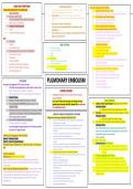
SIGNS AND SYMPTOMS PATHOPHYSIOLOGY Care for a patient with PE (BOX):
peep
PULMONARY EMBOLISM KEY FEATURES (BOX) § Collection of particulate matter (solid, liquid or air) that enters venous § Apply O2 by nasal cannula or mask
§ Classic symptoms circulation and lodge in the pulmonary vessels.
§ Reassure patient that the correct measures are being taken
§ Dyspnea, sudden onset § Large emboli: Obstruct blood flow, reduce gas exchange, reduce
§ Place patient in high fowlers position
oxygenation, pulmonary tissue hypoxia, decreased perfusion and potential
§ Sharp, stabbing chest pain (pleuritic chest pain) death. § Apply telemetry monitoring equipment
§ Apprehension, restlessness § Most common substance is blood clot § Obtain an adequate venous access
§ Feeling of impending doom § It is most common preventable death in hospital but is often § Assess oxygen saturation continuously
§ Cough misdiagnosed § Assess respiratory status at least Q30 min
§ Hemoptysis (bloody sputum) § PE most often occur due to a Venous thromboembolism (VTE) aka - listening to lung sounds (abnormal or crackles)
Deep vein thrombosis DVT. - measuring the rate, rhythm, and ease of respirations
Signs - checking skin color and capillary refill
§ Tachypnea - checking position of trachea
§ Crackles WHO IS AT RISK? § Assess cardiac status by:
§ Pleural friction rub § Prolonged immobility - comparing BP in rt and lt arms
§ Tachycardia CAUSE OF PE FOR PT ON BED REST § Central venous catheters - checking pulse for quality
§ Surgery - checking cardiac monitor for dysrhythmias
§ S3 or S4 heart sounds
§ Obesity - checking for JVD
§ Diaphoresis § Advanced age
§ Ensure that prescribed chest imaging and laboratory tests are
§ Fever (low grade) § Conditions that increase blood clotting
Hx of thromboembolism
obtained immediately
§ Petechiae over the chest and axillae §
§ Smoking, pregnancy, estrogen therapy, § CBC with diff, platelet count, PT, PTT, D-dimer and ABG
§ Decreased arterial oxygen saturations
§ HF, stroke, cancer (lung & prostate) and trauma § Examine the thorax for presence of petechia
§ Systemic hypotension § Administer prescribed anticoagulants
§ Enoxaparin (Lovenox) or Fondaparinux (Arixtra)
PULMONARY EMBOLISM
§ Monitor PTT levels with these drugs (1.5-2.5)
TREATMENT § Assess for bleeding
Nonsurgical management of PE is most common. § Handle patient gently
§ Fibrinolytic drugs (alteplase) is used if patient meets criteria § Institute bleeding precautions
§ Shock, hemodynamic collapse, or instability COMPLICATIONS
§ Must be administered in ICU due to high risk for bleeding • Deoxygenated blood moves into the arterial circulation
§ Heparin therapy causing HYPOXIA
§ Started in hospital usually continues for 5-10 days § Pts w/ s/s of PE who have risk factors and present with JVD, LABS / DIAGNOSTICS
§ Oral anticoagulant WARFARIN started on day 1 or 2 of hypotension and cyanosis call RAPID, elevate HOB and administer § Initially pt will be Hyperventilation triggered by hypoxia leads to
heparin therapy O2. CARDIOGENIC SHOCK respiratory alkalosis
§ Therapy continuous with both heparin and warfarin until INR o Measured by ABG PaCo2 levels decrease - initially
reaches 2.0 to 3.0. Heparin is D/C once INR is greater than 2. § Then later it starts to shunt (blood not getting oxygen) resulting in
PRIORITY NURSING INTERVENTIONS/ TEACHING
§ Other drugs used than warfarin Respiratory acidosis
Preventions of Pulmonary Embolism (BOX) Chart 32-1
o Measured by ABG PaCo2 levels increase
§ Low-molecular weight heparin (enoxaparin) § Passive and active ROM exercises for postop and immobilized patients
Ambulate soon after surgery § Then due to lactic acid build up (tissue hypoxia) resulting in
§ Thrombin inhibitor (rivaroxaban) §
§ Anti-embolism and pneumatic compression stockings after surgery Metabolic acidosis
§ Oral anticoagulant usage continues for 3-6 weeks but others
§ Evaluate the patient for the need of anticoagulants (heparin or § Even if ABG studies and pulse oximetry show hypoxemia, the
may take it for longer periods Enoxaparin) results are not enough alone for diagnosing PE. A patient with a
§ Avoid the use of tight garters, girdles and constricting clothing small embolus may not be hypoxemic.
Antidotes for Anticoagulants and fibrinolytics § Prevent pressure under the popliteal space
§ Other Lab values
Heparin - Protamine sulfate § Do not place a pillow under the knee; instead use alternating pressure
§ BMP, troponin, BNP
Warfarin - Vitamin K1 (phytonadione) mattress
Perform a comprehensive assessment of peripheral circulation § D-dimer test rises with fibrinolysis
Fibrinolytic drugs - Clotting factors - Fresh froze plasma - Aminocaproic §
§ Elevate the affected limb 20 degrees or more above the level of the heart o When the value is normal or low it can rule out PE
acid
improve venous return o If elevated other test are needed to determine a PE
§ Change patient position Q2hrs or ambulate as tolerated occurred
Surgical management § Prevent injury to the vessel lumen by prevention local pressure, trauma, § Pulmonary angiography Best indicator
Embolectomy - Removal of the embolus infection or sepsis
§ Computed tomography pulmonary angiography (CT-PA)
May be performed when fibrinolytic therapy cannot be used for a § Refrain from massaging leg muscle
patient who has massive or multiple emboli with shock or bleeding § Instruct patient not to cross legs § Ventilation perfusion (V/Q) scans not used as much only for
complications § Administer prescribed prophylactic low dose anticoagulant and certain circumstances. Used if pt has Allergies to contrast dye
Inferior vena cava filtration placement of filter - Prevents further emboli antiplatelet drugs § Chest X-ray - May diagnose other conditions that mimic acute PE
§ Teach patient to avoid activities that result in the Valsalva maneuver § Doppler ultrasound - Can verify the presence of DVT and support
from reaching the lungs in patients with ongoing risk for PE. Less risky
§ Breath holding, bearing down for bowel movements, coughing a diagnosis of PE
than drug therapy. Considered for those with: Recurrent or major § Administer prescribed drugs, such as stool softeners
bleeding while receiving anticoagulants. Septic PE. Pulmonary § Prevent episodes of Valsalva maneuver
embolectomy § Teach the patient and family about precautions
§ Encourage smoking cessation
, Pulmonary embolism (PE) severity and management options (BOX)
Prevention of injury for patient on (anticoagulants, Fibrinolytic and Antiplatelets) BOX
Massive PE.- High mortality rate 65%
§ Handle the patient gently Possible symptoms
§
§ Use and teach UAP to use a lift sheet when moving and positioning patient in bed
- Severe hypotension SBP <90 for more than 15min
§ Avoid IM injections and venipunctures
- Cardiac arrest/cardiopulmonary collapse
§ When injections or venipunctures are necessary, use small gauge needles
- Severe bradycardia
§ Apply firm pressure to the needle stick for 10 min or until the site no longer oozes blood
- Shock
§ Apply ice to areas of trauma
- Severe dyspnea/respiratory distress
§ Test all urine, vomit and stool for occult blood
§ Assess IV sites at least every 4 hours for bleeding Management options
§
§ Notify nursing personnel immediately if any trauma occurs and if bleeding or bruising is noted Unfractionated heparin (initial treatment)
-
§ Avoid trauma to rectal tissue
- CPR
§ Do not administer enemas Inotropic or vasopressor support; fluids (Hypotension)
-
§ If suppositories are prescribed, lubricate and administer with caution Dobutamine, norepinephrine/dopamine, crystalloid solution
-
§ Instruct the patient and UAP to use an electric shaver rather than a razor Monitor closely for pulmonary hypertension
-
§ When providing mouth care or supervising others in providing mouth care
- Fibrinolytic therapy
§ Use a soft-bristled toothbrush or tooth sponge
- Tissue plasminogen activator (tPA)
§ Do not use floss
- Alteplase (activase)
§ Check to make certain that dentures fit and do not rub
§ Instruct the patient not to blow the nose forcefully or insert objects into the nose Sub massive PE
§ Ensure that the patient wears shoes with firm soles whenever he or she is ambulating § Possible symptoms
§ Ensure that antidotes to anticoagulation therapy are on unit - Normotension
- RV dysfunction on Echo
- RV dilation on Echo or CT
- Right bundle branch block
Preventing injury and bleeding (BOX) - ST elevation or depression
- T-wave inversion
During the time you are taking anticoagulants - Elevated BNP or troponin
§ Use an electric shaver
§ Use a soft bristle toothbrush and do not floss § Management option
§ Do not have dental health work without consulting with HCP - Low-molecular weight (preferred)
§ Do not take aspirin or salicylate products - Thrombolytic therapy if elevation in troponin levels or BNP
§ Read the label - Fondaparinux (Arixtra)
§ Do not participate in contact sports or any activity that is likely to result in being bumped, scratched and scraped
- Unfractionated heparin
§ If you are bumped, apply ice to the site for at least 1 hour
§ Avoid hard foods that would scrape the inside of your mouth Low-risk PE - Mortality rate is low 1%-8%
§ Eat warm, cool or cold foods to avoid burning your mouth
§ Check your skin and mouth daily for bruises, swelling or areas with small, reddish-purple marks that may indicate § Possible symptoms
bleeding
- Normotension
§ Notify your HCP if you:
- No RV dysfunction
§ Are injured and persistent bleeding results
- No elevation in BNP or troponin
§ Have excessive menstrual bleeding
§ See blood in your urine or bowel movements
§ Avoid anal intercourse § Management option
§ Take a stool softener to prevent straining during a bowel movement - Low-molecular weight (preferred)
§ Do not use enemas or rectal suppositories - Rivaroxaban (Xarelto)
§ Do not wear clothing or shoes that are tight or that rub - Thrombolytics not used due to risk for bleeding
§ Avoid blowing your nose forcefully or placing objects in your nose
§ If you must blow your nose do so gently without blocking either nasal passage
§ Avoid playing musical instruments that raise the pressure inside your head, such as brass wind instruments and
woodwinds or reed instruments.
§ Keep all appointments for laboratory test
§ Monitor for bleeding Q2hrs
§ Measure abdominal girth Q8hrs
§ Reassure the patient to decrease anxiety
, Home care assessment (patient after PE) (BOX)
- Assess respiratory status
o Observe rate and depth of ventilation
o Auscultate lungs
o Examine nail beds and mucous membranes for evidence of cyanosis, indicating reduced
gas exchange
o Take a pulse oximetry reading
o Ask the patient if chest pain or SOB is experienced in any position
o Ask the patient about the presence of sputum and its color and character
- Assess cardiovascular status
o Take vital signs, including apical pulse, pulse pressure
o Note presence or absence of peripheral edema
o Examine hand vein filling in the dependent position
o Examine neck vein filling in the recumbent and sitting position
- Assess lower extremities for DVT
o Examine lower legs and compared with each other for
§ General edema
§ Calf swelling
§ Surface temperature
§ Presence of red streaks or cordlike, palpable structure
o Measure calf circumference
- Assess for evidence of bleeding
o Examine the mouth and gums for oozing or frank bleeding
o Examine all skin areas, especially old puncture sites and wounds, for bleeding, bruising
or petechiae
o If the patient voids during the visit, test the urine for occult blood
- Assess cognition and mental status
o Check LOC
o Check orientation to time, place and person
o Can the patient accurately read a seven-word sentence containing no words with more
Normal Lab Ranges (BOX) than 3 syllables
- Assess the patients understanding of illness and adherence to treatment
o Symptoms to report to HCP
- Partial thromboplastin time (PTT) o Drug therapy plan (correct time and dose)
o Monitor for HEPARIN o Bleeding precautions
o Normal 30-40seconds o Prevention of venous thromboembolism AKA (DVT)
o Therapeutic 1.5-2.5 times the normal
- Prothrombin Time (PT)
o Monitor for WARFARIN
o Normal 11-12.5
o Therapeutic 1.5-2.0 times the normal
- International normalized ratio (INR)
o Monitor for WARFARIN
o Normal 0.8-1.1
o Therapeutic 2.5-3.0 Alert
o Recurrent PE therapeutic 3.0-4.5 - Heparin comes in variety of concentrations in vials that have differing amounts, which contributed to
possible medication errors.
Alert
- Monitor patients at risk to recognize signs and symptoms of PE (SOB, chest pain, hypotension without
any obvious cause). If such symptoms are present, respond by notifying the rapid response team. If PE
is strongly suspected, prompt categorization and management strategies are started before diagnostic
studies have been completed.
, SIGNS AND SYMPTOMS
PATHOPHYSIOLOGY
Oxygenation Failure (hypoxemic) - O2 IS NOT ABLE TO ATTACH TO HEMOGLOBIN ASSESSMENT
o Air movement (ventilation) is normal but blood flow
(perfusion) is decreased § Mismatch with ventilation or perfusion in the § Symptoms of ARF are related to the systemic effects of (Hypoxia,
o Applying 100% O2 does not correct the problem lungs causing a decrease in gas exchange Hypercapnia and acidosis.
o PaO2 <60 mm Hg § Dyspnea is hallmark sign of respiratory failure
§ Normal is 80-100 mm Hg § Acute respiratory failure (ARF) can be either: - Evaluate how breathless the patient becomes while performing
o SaO2 <90% - Ventilatory failure common task
§ Normal is 95-100% - More intense when dyspnea develops rapidly
- Oxygenation failure low - PaO2= arterial oxygen
§ Slowly progressive respiratory failure may be noticed as dyspnea
Common causes of Oxygenation Failure (BOX) blood decrease to the lungs
§ Low atmospheric oxygen concentration
level less than 60 on exertion or lying down (orthopnea) and may find it easier to
High altitude, closed spaces, smoke inhalation, carbon monoxide - Combination of the two breath in upright position
poisoning, pneumonia, CHF with pulmonary edema, pulmonary - Chronic respiratory problems an increase in dyspnea may
embolism, ARDS, interstitial pneumonitis-fibrosis, abnormal represent severe gas exchange problems
hemoglobin, hypovolemic shock, hypoventilation
§ Assess for changes in respiratory rate/patterns and changes in
PT WILL BE lung sounds
§ Complications of nitroprusside (vasodilator) therapy
Thiocyanate toxicity and methemoglobinemia
-dyspnea, restlessness, irritability, confusion, tachycardia, § Monitor ABGs for hypoxia and hypercarbia
decreased LOC, lethargy § Hypoxia respiratory failure symptoms include:
- Restlessness, irritability, agitation, confusion and tachycardia
Ventilatory failure (Hypercapnic) – UNABLE TO TAKE DEEP BREATHS
Blood flow (perfusion) is normal but air movement (ventilation) is inadequate
keep PaO2 >60mm - Oxygen will not fix the problem
o PaCo2 >45 mm Hg -treat the cause § Hypercapnic respiratory failure symptoms include:
o PaCo2 = arterial carbon dioxide more than 45 -oxygen therapy - Decreased LOC, HA, drowsiness, lethargic and seizures
§ Normal is 35-45 mm Hg - Respiratory Acidosis can occur leading to:
o pH < 7.35 - Decreased LOC, drowsiness, confusion, hypotension, bradycardia
§ Normal 7.35-7.45 and weak peripheral pulses.
o SaO2 <90%
Normal is 95-100%
ACUTE RESPIRATORY FAILURE
§
Common causes of Ventilatory Failure (BOX) ventilation is compromised
Extrapulmonary causes:
• Neuromuscular disorders:
o Myasthenia gravis, Guillain-Barre syndrome, LABS / DIAGNOSTICS
poliomyelitis, spinal cord injuries affecting nerves to
intercostal muscles
• Central nervous system dysfunction: COMPLICATIONS
o Stroke, ICP, meningitis
• Chemical depression:
o Opioid analgesics, sedatives, anesthetics, kyphoscoliosis,
massive obesity, sleep apnea, external
obstruction/constriction
Intrapulmonary causes:
• Airway disease:
o COPD and Asthma
• Ventilation-perfusion mismatch:
o Pulmonary embolism, pneumothorax, ARDS, amyloidosis,
pulmonary edema, interstitial fibrosis PRIORITY NURSING INTERVENTIONS/ TEACHING
§ Oxygen therapy is appropriate for any patient with acute
hypoxemia
The patient in ACUTE RESPPIRATORY FAILURE is always hypoxemic caused by an
underlying problem o Used in ARF to keep the PaO2 levels >60 mm Hg
while treating the cause of ARF.
§ If O2 does not maintain acceptable PaO2 levels, Mechanical
TREATMENT
ventilation may be needed
DRUG THERAPY
§ Help the patient find a position of comfort for easier
Nebulizer or Metered dose inhaler (MDI)
breathing
• Dilate the bronchioles and decrease inflammation to
promote gas exchange
o Upright position (fowlers)
• Corticosteroids may be used but benefits have not been § Decrease anxiety caused by dyspnea
demonstrated conclusively o Relaxation techniques, diversion and guided
• Analgesics for pain imagery
§ Encourage deep breathing exercises AND COUGHING
If mechanical ventilation is required
Neuromuscular blockade drugs are prescribed for optimal ventilation COPD PT DIE AT NIGHT AND ALONE
effect
peep
PULMONARY EMBOLISM KEY FEATURES (BOX) § Collection of particulate matter (solid, liquid or air) that enters venous § Apply O2 by nasal cannula or mask
§ Classic symptoms circulation and lodge in the pulmonary vessels.
§ Reassure patient that the correct measures are being taken
§ Dyspnea, sudden onset § Large emboli: Obstruct blood flow, reduce gas exchange, reduce
§ Place patient in high fowlers position
oxygenation, pulmonary tissue hypoxia, decreased perfusion and potential
§ Sharp, stabbing chest pain (pleuritic chest pain) death. § Apply telemetry monitoring equipment
§ Apprehension, restlessness § Most common substance is blood clot § Obtain an adequate venous access
§ Feeling of impending doom § It is most common preventable death in hospital but is often § Assess oxygen saturation continuously
§ Cough misdiagnosed § Assess respiratory status at least Q30 min
§ Hemoptysis (bloody sputum) § PE most often occur due to a Venous thromboembolism (VTE) aka - listening to lung sounds (abnormal or crackles)
Deep vein thrombosis DVT. - measuring the rate, rhythm, and ease of respirations
Signs - checking skin color and capillary refill
§ Tachypnea - checking position of trachea
§ Crackles WHO IS AT RISK? § Assess cardiac status by:
§ Pleural friction rub § Prolonged immobility - comparing BP in rt and lt arms
§ Tachycardia CAUSE OF PE FOR PT ON BED REST § Central venous catheters - checking pulse for quality
§ Surgery - checking cardiac monitor for dysrhythmias
§ S3 or S4 heart sounds
§ Obesity - checking for JVD
§ Diaphoresis § Advanced age
§ Ensure that prescribed chest imaging and laboratory tests are
§ Fever (low grade) § Conditions that increase blood clotting
Hx of thromboembolism
obtained immediately
§ Petechiae over the chest and axillae §
§ Smoking, pregnancy, estrogen therapy, § CBC with diff, platelet count, PT, PTT, D-dimer and ABG
§ Decreased arterial oxygen saturations
§ HF, stroke, cancer (lung & prostate) and trauma § Examine the thorax for presence of petechia
§ Systemic hypotension § Administer prescribed anticoagulants
§ Enoxaparin (Lovenox) or Fondaparinux (Arixtra)
PULMONARY EMBOLISM
§ Monitor PTT levels with these drugs (1.5-2.5)
TREATMENT § Assess for bleeding
Nonsurgical management of PE is most common. § Handle patient gently
§ Fibrinolytic drugs (alteplase) is used if patient meets criteria § Institute bleeding precautions
§ Shock, hemodynamic collapse, or instability COMPLICATIONS
§ Must be administered in ICU due to high risk for bleeding • Deoxygenated blood moves into the arterial circulation
§ Heparin therapy causing HYPOXIA
§ Started in hospital usually continues for 5-10 days § Pts w/ s/s of PE who have risk factors and present with JVD, LABS / DIAGNOSTICS
§ Oral anticoagulant WARFARIN started on day 1 or 2 of hypotension and cyanosis call RAPID, elevate HOB and administer § Initially pt will be Hyperventilation triggered by hypoxia leads to
heparin therapy O2. CARDIOGENIC SHOCK respiratory alkalosis
§ Therapy continuous with both heparin and warfarin until INR o Measured by ABG PaCo2 levels decrease - initially
reaches 2.0 to 3.0. Heparin is D/C once INR is greater than 2. § Then later it starts to shunt (blood not getting oxygen) resulting in
PRIORITY NURSING INTERVENTIONS/ TEACHING
§ Other drugs used than warfarin Respiratory acidosis
Preventions of Pulmonary Embolism (BOX) Chart 32-1
o Measured by ABG PaCo2 levels increase
§ Low-molecular weight heparin (enoxaparin) § Passive and active ROM exercises for postop and immobilized patients
Ambulate soon after surgery § Then due to lactic acid build up (tissue hypoxia) resulting in
§ Thrombin inhibitor (rivaroxaban) §
§ Anti-embolism and pneumatic compression stockings after surgery Metabolic acidosis
§ Oral anticoagulant usage continues for 3-6 weeks but others
§ Evaluate the patient for the need of anticoagulants (heparin or § Even if ABG studies and pulse oximetry show hypoxemia, the
may take it for longer periods Enoxaparin) results are not enough alone for diagnosing PE. A patient with a
§ Avoid the use of tight garters, girdles and constricting clothing small embolus may not be hypoxemic.
Antidotes for Anticoagulants and fibrinolytics § Prevent pressure under the popliteal space
§ Other Lab values
Heparin - Protamine sulfate § Do not place a pillow under the knee; instead use alternating pressure
§ BMP, troponin, BNP
Warfarin - Vitamin K1 (phytonadione) mattress
Perform a comprehensive assessment of peripheral circulation § D-dimer test rises with fibrinolysis
Fibrinolytic drugs - Clotting factors - Fresh froze plasma - Aminocaproic §
§ Elevate the affected limb 20 degrees or more above the level of the heart o When the value is normal or low it can rule out PE
acid
improve venous return o If elevated other test are needed to determine a PE
§ Change patient position Q2hrs or ambulate as tolerated occurred
Surgical management § Prevent injury to the vessel lumen by prevention local pressure, trauma, § Pulmonary angiography Best indicator
Embolectomy - Removal of the embolus infection or sepsis
§ Computed tomography pulmonary angiography (CT-PA)
May be performed when fibrinolytic therapy cannot be used for a § Refrain from massaging leg muscle
patient who has massive or multiple emboli with shock or bleeding § Instruct patient not to cross legs § Ventilation perfusion (V/Q) scans not used as much only for
complications § Administer prescribed prophylactic low dose anticoagulant and certain circumstances. Used if pt has Allergies to contrast dye
Inferior vena cava filtration placement of filter - Prevents further emboli antiplatelet drugs § Chest X-ray - May diagnose other conditions that mimic acute PE
§ Teach patient to avoid activities that result in the Valsalva maneuver § Doppler ultrasound - Can verify the presence of DVT and support
from reaching the lungs in patients with ongoing risk for PE. Less risky
§ Breath holding, bearing down for bowel movements, coughing a diagnosis of PE
than drug therapy. Considered for those with: Recurrent or major § Administer prescribed drugs, such as stool softeners
bleeding while receiving anticoagulants. Septic PE. Pulmonary § Prevent episodes of Valsalva maneuver
embolectomy § Teach the patient and family about precautions
§ Encourage smoking cessation
, Pulmonary embolism (PE) severity and management options (BOX)
Prevention of injury for patient on (anticoagulants, Fibrinolytic and Antiplatelets) BOX
Massive PE.- High mortality rate 65%
§ Handle the patient gently Possible symptoms
§
§ Use and teach UAP to use a lift sheet when moving and positioning patient in bed
- Severe hypotension SBP <90 for more than 15min
§ Avoid IM injections and venipunctures
- Cardiac arrest/cardiopulmonary collapse
§ When injections or venipunctures are necessary, use small gauge needles
- Severe bradycardia
§ Apply firm pressure to the needle stick for 10 min or until the site no longer oozes blood
- Shock
§ Apply ice to areas of trauma
- Severe dyspnea/respiratory distress
§ Test all urine, vomit and stool for occult blood
§ Assess IV sites at least every 4 hours for bleeding Management options
§
§ Notify nursing personnel immediately if any trauma occurs and if bleeding or bruising is noted Unfractionated heparin (initial treatment)
-
§ Avoid trauma to rectal tissue
- CPR
§ Do not administer enemas Inotropic or vasopressor support; fluids (Hypotension)
-
§ If suppositories are prescribed, lubricate and administer with caution Dobutamine, norepinephrine/dopamine, crystalloid solution
-
§ Instruct the patient and UAP to use an electric shaver rather than a razor Monitor closely for pulmonary hypertension
-
§ When providing mouth care or supervising others in providing mouth care
- Fibrinolytic therapy
§ Use a soft-bristled toothbrush or tooth sponge
- Tissue plasminogen activator (tPA)
§ Do not use floss
- Alteplase (activase)
§ Check to make certain that dentures fit and do not rub
§ Instruct the patient not to blow the nose forcefully or insert objects into the nose Sub massive PE
§ Ensure that the patient wears shoes with firm soles whenever he or she is ambulating § Possible symptoms
§ Ensure that antidotes to anticoagulation therapy are on unit - Normotension
- RV dysfunction on Echo
- RV dilation on Echo or CT
- Right bundle branch block
Preventing injury and bleeding (BOX) - ST elevation or depression
- T-wave inversion
During the time you are taking anticoagulants - Elevated BNP or troponin
§ Use an electric shaver
§ Use a soft bristle toothbrush and do not floss § Management option
§ Do not have dental health work without consulting with HCP - Low-molecular weight (preferred)
§ Do not take aspirin or salicylate products - Thrombolytic therapy if elevation in troponin levels or BNP
§ Read the label - Fondaparinux (Arixtra)
§ Do not participate in contact sports or any activity that is likely to result in being bumped, scratched and scraped
- Unfractionated heparin
§ If you are bumped, apply ice to the site for at least 1 hour
§ Avoid hard foods that would scrape the inside of your mouth Low-risk PE - Mortality rate is low 1%-8%
§ Eat warm, cool or cold foods to avoid burning your mouth
§ Check your skin and mouth daily for bruises, swelling or areas with small, reddish-purple marks that may indicate § Possible symptoms
bleeding
- Normotension
§ Notify your HCP if you:
- No RV dysfunction
§ Are injured and persistent bleeding results
- No elevation in BNP or troponin
§ Have excessive menstrual bleeding
§ See blood in your urine or bowel movements
§ Avoid anal intercourse § Management option
§ Take a stool softener to prevent straining during a bowel movement - Low-molecular weight (preferred)
§ Do not use enemas or rectal suppositories - Rivaroxaban (Xarelto)
§ Do not wear clothing or shoes that are tight or that rub - Thrombolytics not used due to risk for bleeding
§ Avoid blowing your nose forcefully or placing objects in your nose
§ If you must blow your nose do so gently without blocking either nasal passage
§ Avoid playing musical instruments that raise the pressure inside your head, such as brass wind instruments and
woodwinds or reed instruments.
§ Keep all appointments for laboratory test
§ Monitor for bleeding Q2hrs
§ Measure abdominal girth Q8hrs
§ Reassure the patient to decrease anxiety
, Home care assessment (patient after PE) (BOX)
- Assess respiratory status
o Observe rate and depth of ventilation
o Auscultate lungs
o Examine nail beds and mucous membranes for evidence of cyanosis, indicating reduced
gas exchange
o Take a pulse oximetry reading
o Ask the patient if chest pain or SOB is experienced in any position
o Ask the patient about the presence of sputum and its color and character
- Assess cardiovascular status
o Take vital signs, including apical pulse, pulse pressure
o Note presence or absence of peripheral edema
o Examine hand vein filling in the dependent position
o Examine neck vein filling in the recumbent and sitting position
- Assess lower extremities for DVT
o Examine lower legs and compared with each other for
§ General edema
§ Calf swelling
§ Surface temperature
§ Presence of red streaks or cordlike, palpable structure
o Measure calf circumference
- Assess for evidence of bleeding
o Examine the mouth and gums for oozing or frank bleeding
o Examine all skin areas, especially old puncture sites and wounds, for bleeding, bruising
or petechiae
o If the patient voids during the visit, test the urine for occult blood
- Assess cognition and mental status
o Check LOC
o Check orientation to time, place and person
o Can the patient accurately read a seven-word sentence containing no words with more
Normal Lab Ranges (BOX) than 3 syllables
- Assess the patients understanding of illness and adherence to treatment
o Symptoms to report to HCP
- Partial thromboplastin time (PTT) o Drug therapy plan (correct time and dose)
o Monitor for HEPARIN o Bleeding precautions
o Normal 30-40seconds o Prevention of venous thromboembolism AKA (DVT)
o Therapeutic 1.5-2.5 times the normal
- Prothrombin Time (PT)
o Monitor for WARFARIN
o Normal 11-12.5
o Therapeutic 1.5-2.0 times the normal
- International normalized ratio (INR)
o Monitor for WARFARIN
o Normal 0.8-1.1
o Therapeutic 2.5-3.0 Alert
o Recurrent PE therapeutic 3.0-4.5 - Heparin comes in variety of concentrations in vials that have differing amounts, which contributed to
possible medication errors.
Alert
- Monitor patients at risk to recognize signs and symptoms of PE (SOB, chest pain, hypotension without
any obvious cause). If such symptoms are present, respond by notifying the rapid response team. If PE
is strongly suspected, prompt categorization and management strategies are started before diagnostic
studies have been completed.
, SIGNS AND SYMPTOMS
PATHOPHYSIOLOGY
Oxygenation Failure (hypoxemic) - O2 IS NOT ABLE TO ATTACH TO HEMOGLOBIN ASSESSMENT
o Air movement (ventilation) is normal but blood flow
(perfusion) is decreased § Mismatch with ventilation or perfusion in the § Symptoms of ARF are related to the systemic effects of (Hypoxia,
o Applying 100% O2 does not correct the problem lungs causing a decrease in gas exchange Hypercapnia and acidosis.
o PaO2 <60 mm Hg § Dyspnea is hallmark sign of respiratory failure
§ Normal is 80-100 mm Hg § Acute respiratory failure (ARF) can be either: - Evaluate how breathless the patient becomes while performing
o SaO2 <90% - Ventilatory failure common task
§ Normal is 95-100% - More intense when dyspnea develops rapidly
- Oxygenation failure low - PaO2= arterial oxygen
§ Slowly progressive respiratory failure may be noticed as dyspnea
Common causes of Oxygenation Failure (BOX) blood decrease to the lungs
§ Low atmospheric oxygen concentration
level less than 60 on exertion or lying down (orthopnea) and may find it easier to
High altitude, closed spaces, smoke inhalation, carbon monoxide - Combination of the two breath in upright position
poisoning, pneumonia, CHF with pulmonary edema, pulmonary - Chronic respiratory problems an increase in dyspnea may
embolism, ARDS, interstitial pneumonitis-fibrosis, abnormal represent severe gas exchange problems
hemoglobin, hypovolemic shock, hypoventilation
§ Assess for changes in respiratory rate/patterns and changes in
PT WILL BE lung sounds
§ Complications of nitroprusside (vasodilator) therapy
Thiocyanate toxicity and methemoglobinemia
-dyspnea, restlessness, irritability, confusion, tachycardia, § Monitor ABGs for hypoxia and hypercarbia
decreased LOC, lethargy § Hypoxia respiratory failure symptoms include:
- Restlessness, irritability, agitation, confusion and tachycardia
Ventilatory failure (Hypercapnic) – UNABLE TO TAKE DEEP BREATHS
Blood flow (perfusion) is normal but air movement (ventilation) is inadequate
keep PaO2 >60mm - Oxygen will not fix the problem
o PaCo2 >45 mm Hg -treat the cause § Hypercapnic respiratory failure symptoms include:
o PaCo2 = arterial carbon dioxide more than 45 -oxygen therapy - Decreased LOC, HA, drowsiness, lethargic and seizures
§ Normal is 35-45 mm Hg - Respiratory Acidosis can occur leading to:
o pH < 7.35 - Decreased LOC, drowsiness, confusion, hypotension, bradycardia
§ Normal 7.35-7.45 and weak peripheral pulses.
o SaO2 <90%
Normal is 95-100%
ACUTE RESPIRATORY FAILURE
§
Common causes of Ventilatory Failure (BOX) ventilation is compromised
Extrapulmonary causes:
• Neuromuscular disorders:
o Myasthenia gravis, Guillain-Barre syndrome, LABS / DIAGNOSTICS
poliomyelitis, spinal cord injuries affecting nerves to
intercostal muscles
• Central nervous system dysfunction: COMPLICATIONS
o Stroke, ICP, meningitis
• Chemical depression:
o Opioid analgesics, sedatives, anesthetics, kyphoscoliosis,
massive obesity, sleep apnea, external
obstruction/constriction
Intrapulmonary causes:
• Airway disease:
o COPD and Asthma
• Ventilation-perfusion mismatch:
o Pulmonary embolism, pneumothorax, ARDS, amyloidosis,
pulmonary edema, interstitial fibrosis PRIORITY NURSING INTERVENTIONS/ TEACHING
§ Oxygen therapy is appropriate for any patient with acute
hypoxemia
The patient in ACUTE RESPPIRATORY FAILURE is always hypoxemic caused by an
underlying problem o Used in ARF to keep the PaO2 levels >60 mm Hg
while treating the cause of ARF.
§ If O2 does not maintain acceptable PaO2 levels, Mechanical
TREATMENT
ventilation may be needed
DRUG THERAPY
§ Help the patient find a position of comfort for easier
Nebulizer or Metered dose inhaler (MDI)
breathing
• Dilate the bronchioles and decrease inflammation to
promote gas exchange
o Upright position (fowlers)
• Corticosteroids may be used but benefits have not been § Decrease anxiety caused by dyspnea
demonstrated conclusively o Relaxation techniques, diversion and guided
• Analgesics for pain imagery
§ Encourage deep breathing exercises AND COUGHING
If mechanical ventilation is required
Neuromuscular blockade drugs are prescribed for optimal ventilation COPD PT DIE AT NIGHT AND ALONE
effect


