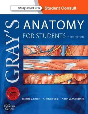
The Elite Universal Test
Bank: Gray's Anatomy
5th Edition
PART 0: THE NAVIGATOR
● PART I: THE PRIMER
○ The "Welcome to the Big Leagues" Hook
○ The "Critical Axioms" Cheat Sheet
● PART II: THE ELITE TEST BANK
○ Tier 1 (Questions 1–28) - Foundational Syntax & Application: The Body, Back,
and Thorax. Testing hard-deck anatomical relationships, cross-sectional imaging,
and cardiopulmonary syntax.
○ Tier 2 (Questions 29–58) - Complex Application & Simulation: The Abdomen,
Pelvis, Perineum, and Lower Limb. Situational variables involving splanchnic
integration, urogenital adaptation, and biomechanical trauma.
○ Tier 3 (Questions 59–88) - Grandmaster Synthesis: The Upper Limb, Head,
Neck, and Neuroanatomy. High-stakes scenarios demanding synthesis of cranial
nerve pathways, brachial plexus crises, and multi-system failures.
PART I: THE PRIMER
Mastering this exhaustive test bank bridges the gap between static anatomical memorization
and the elite, high-stakes clinical intuition required by top-tier global medical boards. By
aggressively intercepting common cognitive traps in neurovascular topography, cross-sectional
relationships, and modern diverse population considerations, you will forge an operational
mastery of the human body that translates directly to diagnostic precision.
The "Critical Axioms" Cheat Sheet
● The Neurovascular Law (Hilton's Law): A nerve crossing a joint invariably innervates
the muscles acting on that joint, the joint capsule itself, and the overlying skin.
● The Fascial Containment Principle: Infections and hemorrhages strictly follow defined
fascial planes; they will not cross compartmental boundaries unless the pressure exceeds
the fascial tensile limit, resulting in compartment syndrome.
● The Referred Pain Axiom: Visceral afferent pain fibers travel retrograde along
sympathetic pathways to the spinal cord; pain is perceived in the somatic dermatome
corresponding to the embryonic origin of the affected organ.
● The "Water Under the Bridge" Mandate: In pelvic surgery, the ureter invariably passes
, inferior and posterior to the uterine artery (or vas deferens); failure to recognize this
guarantees iatrogenic renal failure.
● The Portal-Caval Imperative: In portal hypertension, venous blood bypasses the liver via
critical anastomoses (esophageal, paraumbilical, rectal), forming predictable, lethal
clinical varices.
PART II: THE ELITE TEST BANK
Tier 1 - Foundational Syntax & Application
Q1: An ultrasound of the right upper quadrant is performed using a mid-clavicular sagittal plane.
Based on the principles of radiological anatomy, which structural orientation is the MOST
ACCURATE? A) The superior pole of the kidney appears anterior to the liver edge B) The
gallbladder fundus rests dorsal to the duodenum C) The right hemidiaphragm forms a
hyperechoic boundary superior to the hepatic dome D) The portal vein appears radiolucent
relative to the inferior vena cava
● The Answer: C (The right hemidiaphragm forms a hyperechoic boundary superior to the
hepatic dome)
● Distractor Analysis:
○ A is incorrect: The kidney is retroperitoneal and lies posterior, not anterior, to the
liver edge.
○ B is incorrect: The gallbladder sits in the cystic fossa on the visceral surface of the
liver, ventral to the duodenum.
○ D is incorrect: Radiolucency is a radiographic (X-ray) term; ultrasound utilizes
echogenicity.
The Mentor's Analysis: Cross-sectional imaging relies on absolute positional relationships and
modality-specific terminology. When utilizing ultrasound, tissue density dictates sound wave
reflection. By utilizing anatomical anchors, you bypass the common trap of planar disorientation.
Professional/Academic Intuition: Always define spatial planes using native anatomical
anchors (e.g., diaphragm = superior boundary) and modality-native syntax.
Q2: A patient requires a lumbar puncture. The needle must pass through the ligamentum
flavum. Based on the principles of spinal meningeal architecture, which space must the needle
enter NEXT to sample cerebrospinal fluid? A) Epidural space B) Subdural space C)
Subarachnoid space D) Central canal
● The Answer: C (Subarachnoid space)
● Distractor Analysis:
○ A is incorrect: The epidural space contains fat and venous plexuses, lying
superficial to the dura mater.
○ B is incorrect: The subdural space is a potential space between the dura and
arachnoid, devoid of CSF.
○ D is incorrect: The central canal is within the spinal cord parenchyma, which
terminates at L1/L2.
The Mentor's Analysis: CSF is exclusively housed within the subarachnoid space between the
arachnoid and pia mater. When facing a lumbar puncture, the immediate priority is bypassing
the spinal cord termination. By utilizing the L3/L4 or L4/L5 interspace, you bypass the common
trap of spinal cord trauma. Professional/Academic Intuition: To draw CSF, the needle must
pierce the dura-arachnoid complex below the L2 vertebral level.
, Q3: An MRI reveals a posterolateral herniation of the L4-L5 intervertebral disc. Based on the
principles of lumbar neuroanatomy, which specific nerve root is MOST LIKELY compressed? A)
L3 nerve root B) L4 nerve root C) L5 nerve root D) S1 nerve root
● The Answer: C (L5 nerve root)
● Distractor Analysis:
○ A is incorrect: The L3 root exits far above this structural level.
○ B is incorrect: Due to the downward trajectory of the cauda equina, the L4 root exits
via the superior portion of the L4-L5 foramen, escaping the herniation.
○ D is incorrect: The S1 root remains protected within the dural sac until the L5-S1
level.
The Mentor's Analysis: In the lumbar spine, exiting nerve roots hug the superior pedicle. When
facing a posterolateral herniation, the immediate priority is identifying the traversing root. By
utilizing the descending root logic, you bypass the common trap of matching the exit level to the
disc level. Professional/Academic Intuition: A standard lumbar herniation at level X-Y
compresses traversing root Y.
Q4: A patient undergoes a radical mastectomy, severing the long thoracic nerve. Based on the
principles of thoracic muscular innervation, which physical finding is IMMEDIATELY expected?
A) Inability to initiate arm abduction B) Loss of sensation over the medial arm C) Medial winging
of the scapula upon pushing against a wall D) Weakness in latissimus dorsi extension
● The Answer: C (Medial winging of the scapula upon pushing against a wall)
● Distractor Analysis:
○ A is incorrect: Initiation of abduction is governed by the suprascapular nerve
(supraspinatus).
○ B is incorrect: Sensation over the medial arm is provided by the medial brachial
cutaneous nerve.
○ D is incorrect: The thoracodorsal nerve supplies the latissimus dorsi.
The Mentor's Analysis: The long thoracic nerve strictly innervates the serratus anterior, which
anchors the medial border of the scapula. When facing axillary dissection, the immediate priority
is nerve preservation. By utilizing proper anatomical boundaries, you bypass the common trap
of iatrogenic winging. Professional/Academic Intuition: Medial winging is the absolute clinical
hallmark of long thoracic nerve trauma.
Q5: A central venous line is placed into the right internal jugular vein to rest in the superior vena
cava. Based on the principles of thoracic surface anatomy, which landmark corresponds
EXACTLY to the SVC-atrial junction? A) Clavicular notch B) Xiphisternal joint C) Right 3rd
costal cartilage at the sternal border D) Jugular notch
● The Answer: C (Right 3rd costal cartilage at the sternal border)
● Distractor Analysis:
○ A is incorrect: The clavicular notch marks the sternoclavicular joint, superior to the
SVC.
○ B is incorrect: The xiphisternal joint aligns with the inferior border of the heart (right
ventricle).
○ D is incorrect: The jugular notch marks the T2 vertebral level, far above the right
atrium.
The Mentor's Analysis: Surface anatomy translates internal structures to the chest wall. When
facing central line placement, the immediate priority is identifying the cavoatrial junction. By
utilizing the 3rd costal cartilage, you bypass the common trap of excessively deep catheter
advancement. Professional/Academic Intuition: The superior vena cava enters the right
atrium exactly at the level of the right 3rd costal cartilage.
Bank: Gray's Anatomy
5th Edition
PART 0: THE NAVIGATOR
● PART I: THE PRIMER
○ The "Welcome to the Big Leagues" Hook
○ The "Critical Axioms" Cheat Sheet
● PART II: THE ELITE TEST BANK
○ Tier 1 (Questions 1–28) - Foundational Syntax & Application: The Body, Back,
and Thorax. Testing hard-deck anatomical relationships, cross-sectional imaging,
and cardiopulmonary syntax.
○ Tier 2 (Questions 29–58) - Complex Application & Simulation: The Abdomen,
Pelvis, Perineum, and Lower Limb. Situational variables involving splanchnic
integration, urogenital adaptation, and biomechanical trauma.
○ Tier 3 (Questions 59–88) - Grandmaster Synthesis: The Upper Limb, Head,
Neck, and Neuroanatomy. High-stakes scenarios demanding synthesis of cranial
nerve pathways, brachial plexus crises, and multi-system failures.
PART I: THE PRIMER
Mastering this exhaustive test bank bridges the gap between static anatomical memorization
and the elite, high-stakes clinical intuition required by top-tier global medical boards. By
aggressively intercepting common cognitive traps in neurovascular topography, cross-sectional
relationships, and modern diverse population considerations, you will forge an operational
mastery of the human body that translates directly to diagnostic precision.
The "Critical Axioms" Cheat Sheet
● The Neurovascular Law (Hilton's Law): A nerve crossing a joint invariably innervates
the muscles acting on that joint, the joint capsule itself, and the overlying skin.
● The Fascial Containment Principle: Infections and hemorrhages strictly follow defined
fascial planes; they will not cross compartmental boundaries unless the pressure exceeds
the fascial tensile limit, resulting in compartment syndrome.
● The Referred Pain Axiom: Visceral afferent pain fibers travel retrograde along
sympathetic pathways to the spinal cord; pain is perceived in the somatic dermatome
corresponding to the embryonic origin of the affected organ.
● The "Water Under the Bridge" Mandate: In pelvic surgery, the ureter invariably passes
, inferior and posterior to the uterine artery (or vas deferens); failure to recognize this
guarantees iatrogenic renal failure.
● The Portal-Caval Imperative: In portal hypertension, venous blood bypasses the liver via
critical anastomoses (esophageal, paraumbilical, rectal), forming predictable, lethal
clinical varices.
PART II: THE ELITE TEST BANK
Tier 1 - Foundational Syntax & Application
Q1: An ultrasound of the right upper quadrant is performed using a mid-clavicular sagittal plane.
Based on the principles of radiological anatomy, which structural orientation is the MOST
ACCURATE? A) The superior pole of the kidney appears anterior to the liver edge B) The
gallbladder fundus rests dorsal to the duodenum C) The right hemidiaphragm forms a
hyperechoic boundary superior to the hepatic dome D) The portal vein appears radiolucent
relative to the inferior vena cava
● The Answer: C (The right hemidiaphragm forms a hyperechoic boundary superior to the
hepatic dome)
● Distractor Analysis:
○ A is incorrect: The kidney is retroperitoneal and lies posterior, not anterior, to the
liver edge.
○ B is incorrect: The gallbladder sits in the cystic fossa on the visceral surface of the
liver, ventral to the duodenum.
○ D is incorrect: Radiolucency is a radiographic (X-ray) term; ultrasound utilizes
echogenicity.
The Mentor's Analysis: Cross-sectional imaging relies on absolute positional relationships and
modality-specific terminology. When utilizing ultrasound, tissue density dictates sound wave
reflection. By utilizing anatomical anchors, you bypass the common trap of planar disorientation.
Professional/Academic Intuition: Always define spatial planes using native anatomical
anchors (e.g., diaphragm = superior boundary) and modality-native syntax.
Q2: A patient requires a lumbar puncture. The needle must pass through the ligamentum
flavum. Based on the principles of spinal meningeal architecture, which space must the needle
enter NEXT to sample cerebrospinal fluid? A) Epidural space B) Subdural space C)
Subarachnoid space D) Central canal
● The Answer: C (Subarachnoid space)
● Distractor Analysis:
○ A is incorrect: The epidural space contains fat and venous plexuses, lying
superficial to the dura mater.
○ B is incorrect: The subdural space is a potential space between the dura and
arachnoid, devoid of CSF.
○ D is incorrect: The central canal is within the spinal cord parenchyma, which
terminates at L1/L2.
The Mentor's Analysis: CSF is exclusively housed within the subarachnoid space between the
arachnoid and pia mater. When facing a lumbar puncture, the immediate priority is bypassing
the spinal cord termination. By utilizing the L3/L4 or L4/L5 interspace, you bypass the common
trap of spinal cord trauma. Professional/Academic Intuition: To draw CSF, the needle must
pierce the dura-arachnoid complex below the L2 vertebral level.
, Q3: An MRI reveals a posterolateral herniation of the L4-L5 intervertebral disc. Based on the
principles of lumbar neuroanatomy, which specific nerve root is MOST LIKELY compressed? A)
L3 nerve root B) L4 nerve root C) L5 nerve root D) S1 nerve root
● The Answer: C (L5 nerve root)
● Distractor Analysis:
○ A is incorrect: The L3 root exits far above this structural level.
○ B is incorrect: Due to the downward trajectory of the cauda equina, the L4 root exits
via the superior portion of the L4-L5 foramen, escaping the herniation.
○ D is incorrect: The S1 root remains protected within the dural sac until the L5-S1
level.
The Mentor's Analysis: In the lumbar spine, exiting nerve roots hug the superior pedicle. When
facing a posterolateral herniation, the immediate priority is identifying the traversing root. By
utilizing the descending root logic, you bypass the common trap of matching the exit level to the
disc level. Professional/Academic Intuition: A standard lumbar herniation at level X-Y
compresses traversing root Y.
Q4: A patient undergoes a radical mastectomy, severing the long thoracic nerve. Based on the
principles of thoracic muscular innervation, which physical finding is IMMEDIATELY expected?
A) Inability to initiate arm abduction B) Loss of sensation over the medial arm C) Medial winging
of the scapula upon pushing against a wall D) Weakness in latissimus dorsi extension
● The Answer: C (Medial winging of the scapula upon pushing against a wall)
● Distractor Analysis:
○ A is incorrect: Initiation of abduction is governed by the suprascapular nerve
(supraspinatus).
○ B is incorrect: Sensation over the medial arm is provided by the medial brachial
cutaneous nerve.
○ D is incorrect: The thoracodorsal nerve supplies the latissimus dorsi.
The Mentor's Analysis: The long thoracic nerve strictly innervates the serratus anterior, which
anchors the medial border of the scapula. When facing axillary dissection, the immediate priority
is nerve preservation. By utilizing proper anatomical boundaries, you bypass the common trap
of iatrogenic winging. Professional/Academic Intuition: Medial winging is the absolute clinical
hallmark of long thoracic nerve trauma.
Q5: A central venous line is placed into the right internal jugular vein to rest in the superior vena
cava. Based on the principles of thoracic surface anatomy, which landmark corresponds
EXACTLY to the SVC-atrial junction? A) Clavicular notch B) Xiphisternal joint C) Right 3rd
costal cartilage at the sternal border D) Jugular notch
● The Answer: C (Right 3rd costal cartilage at the sternal border)
● Distractor Analysis:
○ A is incorrect: The clavicular notch marks the sternoclavicular joint, superior to the
SVC.
○ B is incorrect: The xiphisternal joint aligns with the inferior border of the heart (right
ventricle).
○ D is incorrect: The jugular notch marks the T2 vertebral level, far above the right
atrium.
The Mentor's Analysis: Surface anatomy translates internal structures to the chest wall. When
facing central line placement, the immediate priority is identifying the cavoatrial junction. By
utilizing the 3rd costal cartilage, you bypass the common trap of excessively deep catheter
advancement. Professional/Academic Intuition: The superior vena cava enters the right
atrium exactly at the level of the right 3rd costal cartilage.