The ECG and cardiac arrhythmias (hartritmestoornissen)
Heartbeat
Cardiac muscle cells involved in heartbeat;
1. Autorhythmic (cardiac muscle contract on its own without neural or hormonal
stimulation) cells (pacemaker and conducting cells); controls and coordinate
the heartbeat
Pacemaker and conduction cells initiate and distributes electrical signals:
Pacemaker cells (nodal cells);
Establish (ontwikkelen) the normal heart rate
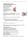
o SA node (cardiac pacemaker); in posterior wall of right atrium.
Driver of the heart rate.
o AV node; between junction atria and ventricles
Membranes of SA and AV nodes have no stable resting. After
repolarization the cell immediately depolarize = pacemaker potential.
Result from slow inflow of Na without compensating outflow of K.
The AV node reaches threshold first it
establishes the basic heart rhythm or
sinus rhythm.
Velocity of the depolarization;
1. SA node
2. AV node
3. AV bundle (bundle of His) , bundle
branches (left and right) and
purkinje fibers (left and right)
Impulse slow as it leaves the internodal pathway and enter the AV node
because nodal cells are smaller than the conducting cells.
So, it takes long to pass through the AV node this allows atria to
contract before ventricles do.
Conducting cells;
Interconnect SA and AV node and distribute contractile stimulus
throughout the myocardium
o Artria; Internodal pathways; distribute the contractile stimulus to
atrial muscle cells
o Ventricle; AV bundle (bundle of His), bundle branches and
purkinje fibers; distribute contractile stimulus to ventricular
myocardium
2. Contractile cells; produce the powerful contraction,
they are the bulk of atrial and ventricular walls
Receive stimulus from purkinje fibers
The cells are interconnected by intercalated discs;
transfer the force from cell to cell and propagate
action potential
The interlocking membranes of adjacent cells held
together by desmosomes and linked by gap
junctions
, Desmosomes; prevent cells from separating during contraction
Gap junctions: allow ions to pass
Action potential in cardiac contractile cells
Resting membrane potential is -90mV comparable to muscle fiber
Action potential in heart takes longer than in muscle
There has to be an action potential (blue) before they contract
(yellow)
Start when membrane of ventricular contractile cell reaches
threshold.
1. Rapid depolarization;
a. Start; Na channel opens (Na flows in) for only a short time = fast
sodium channels.
b. Stop; Na channel close
2. Plateau;
a. Start; Ca channels will open (Ca flows in) for
a long time = slow calcium channels. The
voltage will stay around 0.
b. Stop; Ca channels will close
3. Repolarization;
a. Start; K channels will open (K flows out) for a
long time = slow kalium channels
b. Stop; K channels will close
Refractory period; cardiac contractile cell will not respond to second stimulus for
some time after an action potential starts
Absolute refractor period; rapid depolarization and plateau
The membrane cannot respond at all because Na channels are already open
OR closed and inactivated
Relative refractory period; repolarization
Na-channels are closed but can open. The membrane can respond to stimulus
stronger than the normal one.
In muscle fiber; refractory period will end before muscles deliver maximal tension
(spanning) --> muscle contractions can keep onwards
In cardiac contractile cells; refractory period take until relaxation starts --> cardiac
muscle contractions cannot keep onwards
Role of calcium ions
The action potential produces a contraction by the increase of Ca2+ around the
myofibrils;
1. Extracellular Ca2+ that cross the plasma membrane during plateau phase
provide 20% of Ca2+ required for contraction (direct effect)
2. Arrival of this extracellular Ca2+ triggers release of additional Ca2+ from
reserves in sarcoplasmic reticulum (indirect effect) (C.I.C.R = calcium induced
calcium release)
Pathway
Heartbeat
Cardiac muscle cells involved in heartbeat;
1. Autorhythmic (cardiac muscle contract on its own without neural or hormonal
stimulation) cells (pacemaker and conducting cells); controls and coordinate
the heartbeat
Pacemaker and conduction cells initiate and distributes electrical signals:
Pacemaker cells (nodal cells);
Establish (ontwikkelen) the normal heart rate
o SA node (cardiac pacemaker); in posterior wall of right atrium.
Driver of the heart rate.
o AV node; between junction atria and ventricles
Membranes of SA and AV nodes have no stable resting. After
repolarization the cell immediately depolarize = pacemaker potential.
Result from slow inflow of Na without compensating outflow of K.
The AV node reaches threshold first it
establishes the basic heart rhythm or
sinus rhythm.
Velocity of the depolarization;
1. SA node
2. AV node
3. AV bundle (bundle of His) , bundle
branches (left and right) and
purkinje fibers (left and right)
Impulse slow as it leaves the internodal pathway and enter the AV node
because nodal cells are smaller than the conducting cells.
So, it takes long to pass through the AV node this allows atria to
contract before ventricles do.
Conducting cells;
Interconnect SA and AV node and distribute contractile stimulus
throughout the myocardium
o Artria; Internodal pathways; distribute the contractile stimulus to
atrial muscle cells
o Ventricle; AV bundle (bundle of His), bundle branches and
purkinje fibers; distribute contractile stimulus to ventricular
myocardium
2. Contractile cells; produce the powerful contraction,
they are the bulk of atrial and ventricular walls
Receive stimulus from purkinje fibers
The cells are interconnected by intercalated discs;
transfer the force from cell to cell and propagate
action potential
The interlocking membranes of adjacent cells held
together by desmosomes and linked by gap
junctions
, Desmosomes; prevent cells from separating during contraction
Gap junctions: allow ions to pass
Action potential in cardiac contractile cells
Resting membrane potential is -90mV comparable to muscle fiber
Action potential in heart takes longer than in muscle
There has to be an action potential (blue) before they contract
(yellow)
Start when membrane of ventricular contractile cell reaches
threshold.
1. Rapid depolarization;
a. Start; Na channel opens (Na flows in) for only a short time = fast
sodium channels.
b. Stop; Na channel close
2. Plateau;
a. Start; Ca channels will open (Ca flows in) for
a long time = slow calcium channels. The
voltage will stay around 0.
b. Stop; Ca channels will close
3. Repolarization;
a. Start; K channels will open (K flows out) for a
long time = slow kalium channels
b. Stop; K channels will close
Refractory period; cardiac contractile cell will not respond to second stimulus for
some time after an action potential starts
Absolute refractor period; rapid depolarization and plateau
The membrane cannot respond at all because Na channels are already open
OR closed and inactivated
Relative refractory period; repolarization
Na-channels are closed but can open. The membrane can respond to stimulus
stronger than the normal one.
In muscle fiber; refractory period will end before muscles deliver maximal tension
(spanning) --> muscle contractions can keep onwards
In cardiac contractile cells; refractory period take until relaxation starts --> cardiac
muscle contractions cannot keep onwards
Role of calcium ions
The action potential produces a contraction by the increase of Ca2+ around the
myofibrils;
1. Extracellular Ca2+ that cross the plasma membrane during plateau phase
provide 20% of Ca2+ required for contraction (direct effect)
2. Arrival of this extracellular Ca2+ triggers release of additional Ca2+ from
reserves in sarcoplasmic reticulum (indirect effect) (C.I.C.R = calcium induced
calcium release)
Pathway