Lecture 8 Myocardial perfusion in health and
disease
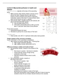
Coronary arteries; originate at the base of the ascending
aorta
1. Right coronary artery (RCA); follows coronary sulcus
around the heart. Supplied blood to right atrium,
portions of both ventricles and portions of electrical
conduction system of the heart most perfusion takes
place during systole and less in diastole
2. Left coronary artery; supplies blood to left ventricle, left
atrium and interventricular septum
a. Left circumflex artery (LCx)
b. Left anterior descending artery (LAD); go to the
apex perfusion takes place during diastole
These blood vessels:
1. Superficial perfusion the heart
2. Penetrate to perfuse the muscle tissue of the heart
Coronary veins;
1. Great cardiac vein (GCV) perfusion takes place during systole
Unique aspects of the coronary circulation
Cyclic compression of the vasculature (vaatstelsel)
1. High resting myocardial metabolic rate: 1 ml/min/g flow
a. High oxygen extraction (60-80%)
b. High capillary density (3000-4000 mm^2)
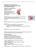
Difference between outside and inside of heart
Sub epicardial vessels: vessels on the surface of the
heart
o Slow blood in systolic + fast blood in diastolic
o Vessel diameter barely change during
contraction of the heart
o Perfusion pressure almost close to aortic
pressure
Sub endocardial vessels: vessels on
the inside of the heart
o Slow blood in systolic + fast
blood in diastolic
o Vessel diameter changes a lot
during contraction of the heart
o Perfusion pressure lower than
in epicardium arterioles due to
resistance of vessels
penetrating from outside to inside of the heart.
How does endocardium cope (omgaan) with this? It tries to lower its
resistance does this by increase vascular density (more vessels)
disease
Coronary arteries; originate at the base of the ascending
aorta
1. Right coronary artery (RCA); follows coronary sulcus
around the heart. Supplied blood to right atrium,
portions of both ventricles and portions of electrical
conduction system of the heart most perfusion takes
place during systole and less in diastole
2. Left coronary artery; supplies blood to left ventricle, left
atrium and interventricular septum
a. Left circumflex artery (LCx)
b. Left anterior descending artery (LAD); go to the
apex perfusion takes place during diastole
These blood vessels:
1. Superficial perfusion the heart
2. Penetrate to perfuse the muscle tissue of the heart
Coronary veins;
1. Great cardiac vein (GCV) perfusion takes place during systole
Unique aspects of the coronary circulation
Cyclic compression of the vasculature (vaatstelsel)
1. High resting myocardial metabolic rate: 1 ml/min/g flow
a. High oxygen extraction (60-80%)
b. High capillary density (3000-4000 mm^2)
Difference between outside and inside of heart
Sub epicardial vessels: vessels on the surface of the
heart
o Slow blood in systolic + fast blood in diastolic
o Vessel diameter barely change during
contraction of the heart
o Perfusion pressure almost close to aortic
pressure
Sub endocardial vessels: vessels on
the inside of the heart
o Slow blood in systolic + fast
blood in diastolic
o Vessel diameter changes a lot
during contraction of the heart
o Perfusion pressure lower than
in epicardium arterioles due to
resistance of vessels
penetrating from outside to inside of the heart.
How does endocardium cope (omgaan) with this? It tries to lower its
resistance does this by increase vascular density (more vessels)