EKG Rhythm Strip Quiz Study Guide (Smarty PANCE Style)
Based on 2026/2027 Syllabus
Instructions: For each strip, identify the rhythm. Choose the single best answer.
Question 1:
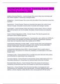
![Strip Description: Regular rhythm, rate ~75 bpm, normal PR interval, normal QRS, one
P wave before each QRS. No extra beats.]
A. Sinus bradycardia
B. Sinus tachycardia
C. Normal sinus rhythm ✓
D. Sinus arrhythmia
ANSWER ✓ Normal sinus rhythm is characterized by a regular rhythm, rate 60-100 bpm,
a constant and normal PR interval, and a P wave before every QRS complex.
Question 2:
![Strip Description: Regular rhythm, rate ~40 bpm, normal P waves, PR, and QRS.]
A. Junctional rhythm
B. Sinus bradycardia ✓
C. Complete heart block
D. Sinus arrest
ANSWER ✓ Sinus bradycardia meets all criteria for normal sinus rhythm except the rate
is less than 60 bpm.
, Question 3:
![Strip Description: Regular rhythm, rate ~130 bpm, normal P waves, PR, and QRS.]
A. Atrial flutter
B. Sinus tachycardia ✓
C. Supraventricular tachycardia (SVT)
D. Ventricular tachycardia
ANSWER ✓ Sinus tachycardia meets all criteria for normal sinus rhythm except the rate
is greater than 100 bpm. P waves are typically upright and normal.
Question 4:
![Strip Description: Rhythm is irregularly irregular. No discernible P waves. Rate ~110
bpm.]
A. Atrial flutter with variable block
B. Sinus arrhythmia
C. Atrial fibrillation ✓
D. Multifocal atrial tachycardia
ANSWER ✓ Atrial fibrillation is classically "irregularly irregular" with no organized P
waves, only fibrillatory waves.
Question 5:
![Strip Description: Sawtooth pattern of atrial flutter waves at ~300/min, ventricular rate
~75 bpm with variable conduction (2:1, 3:1, etc.).]
A. Atrial fibrillation
B. Atrial tachycardia
C. Atrial flutter with variable AV block ✓
D. Sinus rhythm with PACs
ANSWER ✓ Atrial flutter shows characteristic "sawtooth" flutter waves. An irregular
ventricular response indicates variable AV conduction (e.g., 2:1, 3:1, 4:1 block).
, Question 6:
![Strip Description: Regular rhythm, rate ~150 bpm. No visible P waves. QRS is narrow
(<0.12 sec).]
A. Sinus tachycardia
B. Atrial flutter (2:1 block)
C. Supraventricular tachycardia (SVT) ✓
D. Ventricular tachycardia
ANSWER ✓ Supraventricular tachycardia is a regular, rapid rhythm (>150 bpm) with a
narrow QRS complex. P waves are often buried and not visible.
Question 7:
![Strip Description: Regular rhythm, rate ~180 bpm. Wide, bizarre QRS complexes (>0.12
sec). No P waves visible.]
A. SVT with aberrancy
B. Ventricular tachycardia ✓
C. Accelerated idioventricular rhythm
D. Torsades de pointes
ANSWER ✓ Ventricular tachycardia is a regular, rapid rhythm (usually >120 bpm)
originating in the ventricles, characterized by wide, abnormal QRS complexes and often
no associated P waves.
Question 8:
![Strip Description: Irregular rhythm with pauses. A normal sinus beat is followed by an
early, abnormal P wave and a normal QRS. The pause after it is non-compensatory.]
A. Premature ventricular complex (PVC)
B. Premature atrial complex (PAC) ✓
C. Sinus pause
D. Ventricular escape beat
, ANSWER ✓ A PAC is an early beat initiated by an ectopic atrial focus. The P wave looks
different from the sinus P wave, and the following QRS is typically narrow. The
subsequent pause is usually incomplete (non-compensatory).
Question 9:
![Strip Description: An early, wide, and bizarre QRS complex without a preceding P wave.
The T wave is in the opposite direction of the QRS. Followed by a full compensatory
pause.]
A. Premature atrial complex (PAC)
B. Premature junctional complex (PJC)
C. Premature ventricular complex (PVC) ✓
D. Ventricular paced beat
ANSWER ✓ A PVC is characterized by a premature, wide QRS complex (>0.12 sec) that
is bizarre in shape, often with a T wave opposite the QRS vector. The pause following it
is typically a full compensatory pause.
Question 10:
![Strip Description: Regular rhythm, rate ~50 bpm. No P waves before QRS. QRS is
narrow.]
A. Sinus bradycardia
B. Idioventricular rhythm
C. Junctional rhythm ✓
D. Complete heart block
ANSWER ✓ A junctional rhythm (often 40-60 bpm) originates at the AV node. There are
no preceding P waves, or P waves may be inverted and occur after the QRS. The QRS is
typically narrow.
Based on 2026/2027 Syllabus
Instructions: For each strip, identify the rhythm. Choose the single best answer.
Question 1:
![Strip Description: Regular rhythm, rate ~75 bpm, normal PR interval, normal QRS, one
P wave before each QRS. No extra beats.]
A. Sinus bradycardia
B. Sinus tachycardia
C. Normal sinus rhythm ✓
D. Sinus arrhythmia
ANSWER ✓ Normal sinus rhythm is characterized by a regular rhythm, rate 60-100 bpm,
a constant and normal PR interval, and a P wave before every QRS complex.
Question 2:
![Strip Description: Regular rhythm, rate ~40 bpm, normal P waves, PR, and QRS.]
A. Junctional rhythm
B. Sinus bradycardia ✓
C. Complete heart block
D. Sinus arrest
ANSWER ✓ Sinus bradycardia meets all criteria for normal sinus rhythm except the rate
is less than 60 bpm.
, Question 3:
![Strip Description: Regular rhythm, rate ~130 bpm, normal P waves, PR, and QRS.]
A. Atrial flutter
B. Sinus tachycardia ✓
C. Supraventricular tachycardia (SVT)
D. Ventricular tachycardia
ANSWER ✓ Sinus tachycardia meets all criteria for normal sinus rhythm except the rate
is greater than 100 bpm. P waves are typically upright and normal.
Question 4:
![Strip Description: Rhythm is irregularly irregular. No discernible P waves. Rate ~110
bpm.]
A. Atrial flutter with variable block
B. Sinus arrhythmia
C. Atrial fibrillation ✓
D. Multifocal atrial tachycardia
ANSWER ✓ Atrial fibrillation is classically "irregularly irregular" with no organized P
waves, only fibrillatory waves.
Question 5:
![Strip Description: Sawtooth pattern of atrial flutter waves at ~300/min, ventricular rate
~75 bpm with variable conduction (2:1, 3:1, etc.).]
A. Atrial fibrillation
B. Atrial tachycardia
C. Atrial flutter with variable AV block ✓
D. Sinus rhythm with PACs
ANSWER ✓ Atrial flutter shows characteristic "sawtooth" flutter waves. An irregular
ventricular response indicates variable AV conduction (e.g., 2:1, 3:1, 4:1 block).
, Question 6:
![Strip Description: Regular rhythm, rate ~150 bpm. No visible P waves. QRS is narrow
(<0.12 sec).]
A. Sinus tachycardia
B. Atrial flutter (2:1 block)
C. Supraventricular tachycardia (SVT) ✓
D. Ventricular tachycardia
ANSWER ✓ Supraventricular tachycardia is a regular, rapid rhythm (>150 bpm) with a
narrow QRS complex. P waves are often buried and not visible.
Question 7:
![Strip Description: Regular rhythm, rate ~180 bpm. Wide, bizarre QRS complexes (>0.12
sec). No P waves visible.]
A. SVT with aberrancy
B. Ventricular tachycardia ✓
C. Accelerated idioventricular rhythm
D. Torsades de pointes
ANSWER ✓ Ventricular tachycardia is a regular, rapid rhythm (usually >120 bpm)
originating in the ventricles, characterized by wide, abnormal QRS complexes and often
no associated P waves.
Question 8:
![Strip Description: Irregular rhythm with pauses. A normal sinus beat is followed by an
early, abnormal P wave and a normal QRS. The pause after it is non-compensatory.]
A. Premature ventricular complex (PVC)
B. Premature atrial complex (PAC) ✓
C. Sinus pause
D. Ventricular escape beat
, ANSWER ✓ A PAC is an early beat initiated by an ectopic atrial focus. The P wave looks
different from the sinus P wave, and the following QRS is typically narrow. The
subsequent pause is usually incomplete (non-compensatory).
Question 9:
![Strip Description: An early, wide, and bizarre QRS complex without a preceding P wave.
The T wave is in the opposite direction of the QRS. Followed by a full compensatory
pause.]
A. Premature atrial complex (PAC)
B. Premature junctional complex (PJC)
C. Premature ventricular complex (PVC) ✓
D. Ventricular paced beat
ANSWER ✓ A PVC is characterized by a premature, wide QRS complex (>0.12 sec) that
is bizarre in shape, often with a T wave opposite the QRS vector. The pause following it
is typically a full compensatory pause.
Question 10:
![Strip Description: Regular rhythm, rate ~50 bpm. No P waves before QRS. QRS is
narrow.]
A. Sinus bradycardia
B. Idioventricular rhythm
C. Junctional rhythm ✓
D. Complete heart block
ANSWER ✓ A junctional rhythm (often 40-60 bpm) originates at the AV node. There are
no preceding P waves, or P waves may be inverted and occur after the QRS. The QRS is
typically narrow.