Chapter 7: The Skeletal System: The Axial Skeleton
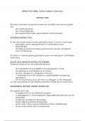
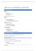
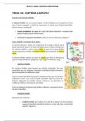
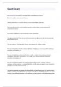
Chapter 7: The Skeletal System: The Axial Skeleton Key Words and Topics Make certain that you can define, and use in context, each of the terms listed below, and that you understand the significance of each of the concepts. 1. Distinguish between the axial and appendicular divisions of the skeleton. • axial skeleton (see the left half of Table 7.1) • appendicular skeleton (see the right half of Table 7.1) The adult human skeleton consists of 206 named bones, most of which are paired, with one member of each pair on the right and left sides of the body. The skeletons of infants and children have more than 206 bones because some of their bones (sacrum and coccyx) fuse later in life. Examples are the hip bones and some bones (sacrum and coccyx) of the vertebral column (backbone). Bones of the adult skeleton are grouped into two principal divisions: the axial skeleton and the appendicular skeleton (appendic‐ = to hang onto). Table 7.1 presents the 80 bones of the axial skeleton and the 126 bones of the appendicular skeleton. Figure 7.1 shows how both divisions join to form the complete skeleton (the bones of the axial skeleton are shown in blue). You can remember the names of the divisions if you think of the axial skeleton as consisting of the bones that lie around the longitudinal axis of the human body, an imaginary vertical line that runs through the body's center of gravity from the head to the space between the feet: skull bones, auditory ossicles (ear bones), hyoid bone (see Figure 7.5), ribs, sternum (breastbone), and bones of the vertebral column. The appendicular skeleton consists of the bones of the upper and lower limbs (extremities or appendages), plus the bones forming the girdles that connect the limbs to the axial skeleton. Functionally, the auditory ossicles in the middle ear, which vibrate in response to sound waves that strike the eardrum, are not part of either the axial or appendicular skeleton, but they are grouped with the axial skeleton for convenience 2. Classify bones on the basis of shape and location. • types of bones Almost all bones of the body can be classified into five main types based on shape: long, short, flat, irregular, and sesamoid (Figure 7.2). As you learned in Chapter 6, long bones have greater length than width, consist of a shaft and a variable number of extremities or epiphyses (ends), and are slightly curved for strength. A curved bone absorbs the stress of the body's weight at several different points, so that it is evenly distributed. If bones were straight, the weight of the body would be unevenly distributed, and the bone would fracture more easily. Long bones consist mostly of compact bone tissue in their diaphyses but have considerable amounts of spongy bone tissue in their epiphyses. Long bones vary tremendously in size and include those in the femur (thigh bone), tibia and fibula (leg bones), humerus (arm bone), ulna and radius (forearm bones), and phalanges (finger and toe bones). long bone long bones have greater length than width, consist of a shaft and a variable number of extremities or epiphyses (ends), and are slightly curved for strength. A curved bone absorbs the stress of the body's weight at several different points, so that it is evenly distributed. If bones were straight, the weight of the body would be unevenly distributed, and the bone would fracture more easily. Long bones consist mostly of compact bone tissue in their diaphyses but have considerable amounts of spongy bone tissue in their epiphyses. Long bones vary tremendously in size and include those in the femur (thigh bone), tibia and fibula (leg bones), humerus (arm bone), ulna and radius (forearm bones), and phalanges (finger and toe bones). short bone are somewhat cube‐shaped and are nearly equal in length and width. They consist of spongy bone tissue except at the surface, which has a thin layer of compact bone tissue. Examples of short bones are most carpal (wrist) bones and most tarsal (ankle) bones. flat bone are generally thin and composed of two nearly parallel plates of compact bone tissue enclosing a layer of spongy bone tissue. Flat bones afford considerable protection and provide extensive areas for muscle attachment. Flat bones include the cranial bones, which protect the brain; the sternum (breastbone) and ribs, which protect organs in the thorax; and the scapulae (shoulder blades). irregular bone have complex shapes and cannot be grouped into any of the previous categories. They vary in the amount of spongy and compact bone present. Such bones include the vertebrae (backbones), hip bones, certain facial bones, and the calcaneus. sesamoid bone (SES‐a‐moyd = shaped like a sesame seed) develop in certain tendons where there is considerable friction, tension, and physical stress, such as the palms and soles. They may vary in number from person to person, are not always completely ossified, and typically measure only a few millimeters in diameter. Notable exceptions are the two patellae (kneecaps), large sesamoid bones located in the quadriceps femoris tendon (see Figure 11.20a) that are normally present in everyone. Functionally, sesamoid bones protect tendons from excessive wear and tear, and they often change the direction of pull of a tendon, which improves the mechanical advantage at a joint. sutural bone An additional type of bone is classified by location rather than shape. Sutural bones (SOO‐chur‐al; sutur‐ = seam) are small bones located in sutures (joints) between certain cranial bones (see Figure 7.6). Their number varies greatly from person to person. 3. Describe the major surface markings on bones and the functions of each. bone surface markings (fissure, foramen, fossa, sulcus, meatus, condyle, facet, head, crest, epicondyle, line, spinous process, trochanter, tubercle, tuberosity) - Bones have characteristic surface markings, structural features adapted for specific functions. Most are not present at birth but develop in response to certain forces and are most prominent in the adult skeleton. In response to tension on a bone surface from tendons, ligaments, aponeuroses, and fasciae, new bone is deposited, resulting in raised or roughened areas. Conversely, compression on a bone surface results in a depression. There are two major types of surface markings: (1) depressions and openings, which allow the passage of soft tissues (such as blood vessels, nerves, ligaments, and tendons) or form joints, and (2)processes, projections or outgrowths that either help form joints or serve as attachment points for connective tissue (such as ligaments and tendons). Table 7.2 describes the various surface markings and provides examples of each. • Use the volume titled “Brief Atlas of the Skeleton, Surface Anatomy, and Selected Medical Images,” which accompanies the textbook, to help you as you study the individual bones. 4. Identify the names, locations and major surface markings of the bones of the skull. • Skull The skull is the bony framework of the head. It contains 22 bones (not counting the bones of the middle ears) and rests on the superior end of the vertebral column (backbone). The bones of the skull are grouped into two categories: cranial bones and facial bones • cranial bones . The cranial bones (crani‐ = brain case) form the cranial cavity, which encloses and protects the brain. The eight cranial bones are the frontal bone, two parietal bones, two temporal bones, the occipital bone, the sphenoid bone, and the ethmoid bone. • • frontal bone- Fourteen facial bones form the face: two nasal bones, two maxillae (or maxillas), two zygomatic bones, the mandible, two lacrimal bones, two palatine bones, two inferior nasal conchae, and the vomer. Exhibits 7.A through 7.G illustrate the bones of the skull from different views. • supraorbital margin- * • parietal bones (2)- * • temporal bones (2)-* • zygomatic arch-* • external auditory meatus-* • mastoid process-* • internal auditory meatus-* • occipital bone* • foramen magnum* • occipital condyles* • sphenoid bone-* • optic foramen-* • ethmoid bone* • superior and middle nasal conchae (turbinates; singular is concha)* • facial bones (see above under “frontal lobe”)* • nasal bones (2)* • maxillae (2; singular is maxilla)* • zygomatic bones (2)* • lacrimal bones (2)* • lacrimal fossa* • palatine bones (2)* • inferior nasal conchae or turbinates (2)* • vomer* • mandible* • body* • rami (2) (singular is ramus)-7G* • angles (2)-7G* • coronoid processes (2)-7G * • condylar processes (2) • temporomandibular joints (2) • alveolar process—on exhibit 7A* • nasal septum* • orbits (2)* Seven bones of the skull join to form each orbit (eye socket) or orbital cavity, which contains the eyeball and associated structures (Figure 7.12). The three cranial bones of the orbit are the frontal, sphenoid, and ethmoid; the four facial bones are the palatine, zygomatic, lacrimal, and maxilla. Each pyramid‐ shaped orbit has four regions that converge posteriorly: 1. Parts of the frontal and sphenoid bones comprise the roof of the orbit. 2. Parts of the zygomatic and sphenoid bones form the lateral wall of the orbit. 3. Parts of the maxilla, zygomatic, and palatine bones make up the floor of the orbit. 4. Parts of the maxilla, lacrimal, ethmoid, and sphenoid bones form the medial wall of the orbit. 5. Identify the principal sutures, sinuses, and fontanels of the skull, as well as the hyoid bone. • suture • coronal suture • sagittal suture • lambdoid suture • squamous sutures (2) The skull exhibits several unique features not seen in other bones of the body. These include sutures, paranasal sinuses, and fontanels. A suture (SOO‐chur = seam) is an immovable joint in most cases in an adult skull that holds most skull bones together. Sutures in the skulls of infants and children, however, often are movable and function as important growth centers in the developing skull. The names of many sutures reflect the bones they unite. For example, the frontozygomatic suture is between the frontal bone and the zygomatic bone. Similarly, the sphenoparietal suture is between the sphenoid bone and the parietal bone. In other cases, however, the names of sutures are not so obvious. Of the many sutures found in the skull, we will identify only four prominent ones: - The coronal suture (KO‐rō‐nal; coron‐ = relating to the frontal or coronal plane) unites the frontal bone and both parietal bones (see Figure 7.4b). - The sagittal suture (SAJ‐i‐tal; sagitt‐ = arrow) unites the two parietal bones on the superior midline of the skull (see Figure 7.4a). The sagittal suture is so named because in the infant, before the bones of the skull are firmly united, the suture and the fontanels (soft spots) associated with it resemble an arrow. - The lambdoid suture (LAM‐doyd) unites the two parietal bones to the occipital bone. This suture is so named because of its resemblance to the capital Greek letter lambda (Λ), as can be seen in Figure 7.6 (with the help of a little imagination). Sutural bones may occur within the sagittal and lambdoid sutures. - The two squamous sutures (SKWĀ‐mus; squam‐ = flat, like the flat overlapping scales of a snake) unite the parietal and temporal bones on the lateral aspects of the skull (see Figure 7.4b). • sinus • paranasal sinus The paranasal sinuses (par′‐a‐NĀ‐zal SĪ‐nus‐ez; para‐ = beside) are cavities within certain cranial and facial bones near the nasal cavity. They are most evident in a sagittal section of the skull (Figure7.13). The paranasal sinuses are lined with mucous membranes that are continuous with the lining of the nasal cavity. Secretions produced by the mucous membranes of the paranasal sinuses drain into the lateral wall of the nasal cavity. Paranasal sinuses are quite small or absent at birth, but increase in size during two periods of facial enlargement—during the eruption of the teeth and at the onset of puberty. They arise as outgrowths of the nasal mucosa that project into the surrounding bones. Skull bones containing the paranasal sinuses are the frontal, sphenoid, ethmoid, and maxillae. The paranasal sinuses allow the skull to increase in size without a change in the mass (weight) of the bone. The paranasal sinuses increase the surface area of the nasal mucosa, thus increasing the production of mucus to help moisten and cleanse inhaled air. In addition, the paranasal sinuses serve as resonating (echo) chambers within the skull that intensify and prolong sounds, thereby enhancing the quality of the voice. The influence of the paranasal sinuses on your voice becomes obvious when you have a cold; the passageways through which sound travels into and out of the paranasal sinuses become blocked by excess mucus production, changing the quality of your voice.
École, étude et sujet
- Établissement
- The Skeletal System
- Cours
- The Skeletal System
Infos sur le Document
- Publié le
- 19 septembre 2023
- Nombre de pages
- 19
- Écrit en
- 2023/2024
- Type
- Examen
- Contient
- Questions et réponses
Sujets
-
chapter 7 the skeletal system the axial skeleton