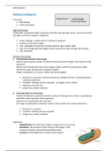
Cell Structure
Methods of studying cells
Two ways: Magnification = size of image
1. Microscopy Size of real object
2. Cell fractionation
Light microscopy
Visible light is passed through a specimen and then through glass lenses, the lenses refract
the light so that the change is magnified.
ü Direct imaging – sample doesn’t need pre-treatment
ü Produces real colour images
ü Fast, adaptable to all kinds of sample systems (gas, liquid, solid)
ü Easy to be integrated with digital camera systems for data storage and analysis
û Low resolution
Electron microscopy
1. Transmission electron microscope
Electron gun produces a beam of electrons which passes through a thin section of the
specimen.
Denser parts absorb electrons and so appear darker, while less dense parts allow
electrons to pass through and so appear brighter.
Image is produced on a screen, called a photomicrograph.
û Must be in a vacuum to prevent electrons colliding with air, so living specimen
cannot be observed
û Complex ‘staining’ process required – so image is not in colour
û Specimen must be thin
û Image may contain artefacts
2. Scanning electron microscope
A beam of electrons is directed onto the surface of the specimen, which is passed back
and forth across a portion of the specimen.
Electrons are scattered by the specimen.
3D image is produced by computer analysis of the pattern of scattered electrons.
û Must be in a vacuum
û Complex ‘staining’ process required
û Image may contain artefacts
Definitions
- Magnification: the ratio of an object’s image size to its real size.
- Resolution: the measure of the clarity of the image, or the
minimum distance of two distinguishable points.
- Contrast: visible differences in parts of the sample.