UCL PHOL1001: Topic 7 - Cardiovascular
System
The action potentials in the heart are about 100 times longer than those of skeletal muscle. - ANS True
The action potential of skeletal muscle is about 2 ms in duration while the action potentials of the heart
cells range from about 150 ms in the cells of the SA node to about 300 ms in a Purkinje fiber.
The cells of the sinoatrial node have a steady resting potential of -90 mV. - ANS False
The membrane potential of the SA node cells is low, about -60 mV, while that of ventricular cells is
about -90 mV.
The cardiac action potential is conducted through the myocardium entirely via specialized conducting
fibres. - ANS False
Although the conduction of the cardiac impulse through the atria occurs preferentially via certain fiber
bundles, these cells are normal atrial myocytes.
The spread of cardiac excitation is delayed by about 0.1 s at the atrioventricular node. - ANS True
The conducting tissue of the heart is composed of specialized cardiac myocytes linked by gap junctions. -
ANS True
In the ventricles, the specialized conducting cells are the bundle cells and the Purkinje fibers, which
transmit their action potentials to ventricular myocytes via gap junctions.
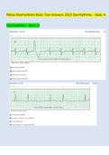
The P wave of the ECG reflects atrial contraction. - ANS False
, The P wave reflects atrial depolarization and precedes atrial contraction.
The QRST complex of the ECG reflects the time during which ventricular fibers are depolarised. - ANS
True
The peak amplitude of the ECG is about 1 mV. - ANS True
The T wave reflects the repolarization of the ventricular fibres. - ANS True
The P-Q interval is normally about 0.1 s. - ANS True
During ventricular diastole the pressure in the left ventricle is close to zero. - ANS True
During ventricular systole, the pressure in the left ventricle reaches a maximum of about 16 kPa (120
mmHg) - ANS True
During ventricular systole, all the blood in the ventricles is ejected. - ANS False
The volume of blood in the ventricles at the end of diastole is about 120 ml. Of this about 70 ml is
ejected (the stroke volume). The ratio of the stroke volume to the end-diastolic volume is called the
ejection fraction and is usually about 60 per cent at rest.
During the initial stage of ventricular contraction the volume of the ventricle does not change. - ANS
True
The mitral valve closes because the pressure in the left ventricle exceeds that in the left atrium. - ANS
True
System
The action potentials in the heart are about 100 times longer than those of skeletal muscle. - ANS True
The action potential of skeletal muscle is about 2 ms in duration while the action potentials of the heart
cells range from about 150 ms in the cells of the SA node to about 300 ms in a Purkinje fiber.
The cells of the sinoatrial node have a steady resting potential of -90 mV. - ANS False
The membrane potential of the SA node cells is low, about -60 mV, while that of ventricular cells is
about -90 mV.
The cardiac action potential is conducted through the myocardium entirely via specialized conducting
fibres. - ANS False
Although the conduction of the cardiac impulse through the atria occurs preferentially via certain fiber
bundles, these cells are normal atrial myocytes.
The spread of cardiac excitation is delayed by about 0.1 s at the atrioventricular node. - ANS True
The conducting tissue of the heart is composed of specialized cardiac myocytes linked by gap junctions. -
ANS True
In the ventricles, the specialized conducting cells are the bundle cells and the Purkinje fibers, which
transmit their action potentials to ventricular myocytes via gap junctions.
The P wave of the ECG reflects atrial contraction. - ANS False
, The P wave reflects atrial depolarization and precedes atrial contraction.
The QRST complex of the ECG reflects the time during which ventricular fibers are depolarised. - ANS
True
The peak amplitude of the ECG is about 1 mV. - ANS True
The T wave reflects the repolarization of the ventricular fibres. - ANS True
The P-Q interval is normally about 0.1 s. - ANS True
During ventricular diastole the pressure in the left ventricle is close to zero. - ANS True
During ventricular systole, the pressure in the left ventricle reaches a maximum of about 16 kPa (120
mmHg) - ANS True
During ventricular systole, all the blood in the ventricles is ejected. - ANS False
The volume of blood in the ventricles at the end of diastole is about 120 ml. Of this about 70 ml is
ejected (the stroke volume). The ratio of the stroke volume to the end-diastolic volume is called the
ejection fraction and is usually about 60 per cent at rest.
During the initial stage of ventricular contraction the volume of the ventricle does not change. - ANS
True
The mitral valve closes because the pressure in the left ventricle exceeds that in the left atrium. - ANS
True