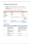
Methods of Studying Cells
Magnification - how many times bigger the image is when compared to the object
Resolution - the minimum distance apart that two objects can be in order for them to be
seen as separate items
Resolving power depends on the wavelength or the form of radiation used
Features Light Electron
Wavelength 400 nm 1 nm
Resolution 200 nm 0.5 nm
Maximum useful x1500 X1,500,000
magnification
Image Natural colour of specimen or dye/ink Black and White
Colour
enhanced for
more detail
Specimens Living or non-living Non-living
Advantages Some organelles bigger than 0.2 um can be High resolution and
seen organelle detail
Types of Microscopes
TEMS SEMS
Electromagnets focus a Scan a beam of electrons across the
beam of electrons which specimen, which knocks electrons off
are transmitted through a the specimen. These are collected in a
specimen cathode tube to form an image
Denser parts of specimen The images show the surface of the
absorb more electrons specimen
which makes them look Can be 3D
darker
High resolution (internal Lower resolution
organelles)
Only thin specimens Can be thick
,Cell Fractionation
To look at organelles under an electron microscope, you need to separate them from the
rest of the cell
1. Homogenisation - breaking up the cell
2. Filtration - getting rid of products you don’t require
3. Ultracentrifugation - separating out the organelles you want by mass (from heaviest to
lightest)
Homogenisation : can be done with two methods
1. Vibrating the cells
2. Placing cells in blender
These methods break up the plasma membrane and release the organelles into solution
To do this, buffer solution is added to maintain the correct pH
(changes in pH could change, denature or affect the functioning of the enzymes)
The solution should be isotonic (same concentration of chemicals as the cells broken down
to prevent damage to organelles by osmosis)
The solution must be ice cold - this stops enzymatic reactions that could break down
organelles
Filtration
The homogenised cell solution (homogenate) is filtered through a gauze to separate the cell
debris from the organelles
The organelles pass through the gauze as they are much smaller than the debris
Ultracentrifugation
Filtration leaves you with a solution containing a mixture of organelles,
Ultracentrifugation is the process by which the fragments in the filtered homogenate are
separated
They are separated in order of mass
Occurs in a centrifugal
1. Low Speed Centrifugation - heaviest organelles forced to bottom of tube and form thin
sediment or pellet
The fluid at the top of the tube (the supernatant) is removed, leaving just the sediment at
the bottom
The supernatant is transferred to another tube in the centrifugal and spun even faster
2. Medium Speed Centrifugation
3. High Speed Centrifugation
4. Very High Speed Centrifugation
The process is continued in this way, so each increase in speed causes the next heaviest
organelle to be sedimented until all the organelles are separated
Eukaryotic Cell Structure
, The Different Organelles:
1. Nucleus
2. Rough endoplasmic reticulum (RER) and Smooth Endoplasmic Reticulum
3. Mitochondria
4. Golgi Apparatus and Golgi Vesicles
5. Lysosomes
6. Ribosomes
7. Cell Wall
8. Chloroplasts
9. Cell Vacuole
In complex multicellular organisms, eukaryotic cells become specialised for specific
functions
Specialised cells are organised into tissues, tissues into organs and organs into
systems
Cell Wall
Structure:
Consists of microfibrils of the polysaccharide cellulose, embedded in a matrix and many
other polysaccharides
Cellulose microfibrils have considerable strength and so contribute to the overall strength of
the cell wall
Middle lamella - a thin layer which marks the boundary between adjacent cell walls and
cements adjacent cells together
Cell walls of algae are made up of cellulose, glycoproteins or both
Cell walls of fungi do not contain cellulose - instead a mixture of chitin (a nitrogen-containing
polysaccharide) and a polysaccharide called glycan and glycoproteins.
Functions:
1. To provide mechanical strength in order to prevent the cell bursting under the pressure
created by the osmotic entry of water
2. Give mechanical strength to the plant as a whole
3. Allow water to pass along it, and so contribute to the movement of water through the plant
Chloroplasts
Structure: 2-10 µm in length and 1 µm in diameter
The Chloroplast Envelope - a double plasma membrane that surrounds the organelle and is
highly selective in what it allows to enter and leave the chloroplast
The Grana- stacks of 100 disc like structures called thylakoids (also where the 1st stage of
photosynthesis takes place-light absorption)
Within the Thylakoids is the photosynthetic pigment called chlorophyll
Some thylakoids have tubular extensions that join up with thylakoids in adjacent grana
The Stroma- a fluid filled matrix, where the second stage of photosynthesis takes place
(synthesis of sugars)
Within the stroma are other structures such as starch grains
Function - harvesting sunlight and carrying out photosynthesis