

The Nervous System
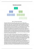
Figure 1: The Nervous System Division
The nervous system is the most complex system within the human body; using a
combination of chemical and electrical signalling between cells to complete thousands of
functions which keep us alive. The nervous system is primarily divided into two areas - the
central nervous system and the peripheral nervous system. The central nervous system
(CNS) is comprised of the brain and the spinal cord - a long bundle of nerve fibres which
extends from the base of the brain to the bottom of the spine protected by the spine. These
make up the control centre of the body, which receives environmental and sensory
information from the peripheral nervous system and returns commands in response to this
information. The peripheral nervous system (PNS) is the network of nerve fibres which
extend from the spinal cord, all across the body. From the brain extends 12 pairs of cranial
nerves which connect it to the sensory organs such as eyes and ears, and to the muscles of
the head and neck. 31 pairs of spinal nerves branch out from the spinal cord to connect it to
the tissues of the limbs, thorax and abdomen (1). These nerves carry signals to the CNS all
the way to the outermost parts of the body.
As seen in figure 1 above, the PNS is comprised of the somatic nervous system and
autonomic nervous system. The somatic nervous system (SNS) is responsible for conscious
actions such as raising one arm while the autonomic nervous system (ANS) is beyond
conscious control, it acts ‘automatically’ to complete functions such as regulating the heart
rate and ventilation. The ANS can be sub-divided into the parasympathetic nervous system
and the sympathetic nervous system. The sympathetic nervous system controls the ‘fight or
flight’ response which occurs when the body is under stress, some actions include
increasing heart rate, dilating bronchia and stimulating adrenaline release. The
parasympathetic nervous system (PSNS) is responsible for the ‘rest and digest’ state which
occurs when the body is not in the fight or flight state. Actions of this system include
reducing heart rate and constricting the bronchia. A third subdivision of the ANS, sometimes
considered a third branch of the peripheral nervous system, is this enteric nervous system.
This system is found within the lining of the gastrointestinal tract and controls solely the
gastrointestinal (GI) processes. The PSNS stimulates the enteric nervous system while the
, sympathetic can inhibit it, meaning the GI system is most active during the rest and digest
state and is mostly inactive during the fight and flight state. Each division of the peripheral
nervous system is made up of two pathways, or ‘arms’, the afferent and the efferent (2). The
afferent arm includes the sensory neurones which connect the receptors – tissues which
detect changes in both the internal and external environment (stimuli) such as temperature –
to the CNS. The efferent arm consists of motor neurones which allow signals to be
transmitted from the CNS to effector organs – muscles and glands which perform an action
in response to stimuli, for example sweat glands producing sweat when a rise in temperature
is detected.
The sensory neurones of the afferent pathway, seen in figure 2 below (left) collect
information about the environment, for example information about light levels is passed
along the optic nerve in the eye to the brain. The axon contains a myelin sheath and extends
from either side of the cell body, these neurons connect receptor cells, such as the
photosensitive retinal cells of the eye, to the CNS. Motor neurones of the efferent pathway
(see figure 2, right) connect the spinal cord to the brain (upper motor neurones) or the
muscles (lower motor neurones) to directly control muscle movement - these neurons have a
myelinated axon connecting the cell body to the effector cells. Relay neurons connect
sensory and motor neurones to one another, their axon is non-myelinated (not surrounded
my myelin sheath) as seen in figure 2 below (middle).
Figure 2: Types of Neurones (25)
The myelin sheath is a lipid-rich substance found surrounding the axons of ‘myelinated’
neurons (3). Regularly occurring gaps in the myelin sheath called the ‘Nodes of Ranvier’
contain the majority of the ion channels of the axon, allowing action potentials to be
generated here – this is essential for nerve signalling. Neurons of the CNS contain
‘oligodendrocyte’ cells which produce myelin – each of these cells can myelinate up to 50
axons. Oligodendrocyte cells are one type of neuroglia – cells with specialised functions that
support the neurons (4). Other types of neuroglia such as microglia, which help protect the
CNS from microorganisms and clean up dead neurones, are also present. In the PNS,
another type of neuroglia, myelin-producing cells known as Schwann cells, myelinate around
100 micrometres of a single axon, each axon requires around 10,000 Schwann cells to
myelinate 1 metre in length. Myelinated axons have two resulting properties which allow
them to conduct an action potential more quickly than non-myelinated axons. Firstly, the high
membrane resistance of the myelin inhibits ion movement in myelinated section, areas other
than the nodes of Ranvier, concentration ions more highly at the nodes to allow rapid
depolarisation and generation of an action potential. Secondly, the reduced capacitance of
the axon lowers the change in ion concentration which is required to generate potential;
these two factors allow for faster signalling through ‘salutatory conduction’, the process in
Figure 1: The Nervous System Division
The nervous system is the most complex system within the human body; using a
combination of chemical and electrical signalling between cells to complete thousands of
functions which keep us alive. The nervous system is primarily divided into two areas - the
central nervous system and the peripheral nervous system. The central nervous system
(CNS) is comprised of the brain and the spinal cord - a long bundle of nerve fibres which
extends from the base of the brain to the bottom of the spine protected by the spine. These
make up the control centre of the body, which receives environmental and sensory
information from the peripheral nervous system and returns commands in response to this
information. The peripheral nervous system (PNS) is the network of nerve fibres which
extend from the spinal cord, all across the body. From the brain extends 12 pairs of cranial
nerves which connect it to the sensory organs such as eyes and ears, and to the muscles of
the head and neck. 31 pairs of spinal nerves branch out from the spinal cord to connect it to
the tissues of the limbs, thorax and abdomen (1). These nerves carry signals to the CNS all
the way to the outermost parts of the body.
As seen in figure 1 above, the PNS is comprised of the somatic nervous system and
autonomic nervous system. The somatic nervous system (SNS) is responsible for conscious
actions such as raising one arm while the autonomic nervous system (ANS) is beyond
conscious control, it acts ‘automatically’ to complete functions such as regulating the heart
rate and ventilation. The ANS can be sub-divided into the parasympathetic nervous system
and the sympathetic nervous system. The sympathetic nervous system controls the ‘fight or
flight’ response which occurs when the body is under stress, some actions include
increasing heart rate, dilating bronchia and stimulating adrenaline release. The
parasympathetic nervous system (PSNS) is responsible for the ‘rest and digest’ state which
occurs when the body is not in the fight or flight state. Actions of this system include
reducing heart rate and constricting the bronchia. A third subdivision of the ANS, sometimes
considered a third branch of the peripheral nervous system, is this enteric nervous system.
This system is found within the lining of the gastrointestinal tract and controls solely the
gastrointestinal (GI) processes. The PSNS stimulates the enteric nervous system while the
, sympathetic can inhibit it, meaning the GI system is most active during the rest and digest
state and is mostly inactive during the fight and flight state. Each division of the peripheral
nervous system is made up of two pathways, or ‘arms’, the afferent and the efferent (2). The
afferent arm includes the sensory neurones which connect the receptors – tissues which
detect changes in both the internal and external environment (stimuli) such as temperature –
to the CNS. The efferent arm consists of motor neurones which allow signals to be
transmitted from the CNS to effector organs – muscles and glands which perform an action
in response to stimuli, for example sweat glands producing sweat when a rise in temperature
is detected.
The sensory neurones of the afferent pathway, seen in figure 2 below (left) collect
information about the environment, for example information about light levels is passed
along the optic nerve in the eye to the brain. The axon contains a myelin sheath and extends
from either side of the cell body, these neurons connect receptor cells, such as the
photosensitive retinal cells of the eye, to the CNS. Motor neurones of the efferent pathway
(see figure 2, right) connect the spinal cord to the brain (upper motor neurones) or the
muscles (lower motor neurones) to directly control muscle movement - these neurons have a
myelinated axon connecting the cell body to the effector cells. Relay neurons connect
sensory and motor neurones to one another, their axon is non-myelinated (not surrounded
my myelin sheath) as seen in figure 2 below (middle).
Figure 2: Types of Neurones (25)
The myelin sheath is a lipid-rich substance found surrounding the axons of ‘myelinated’
neurons (3). Regularly occurring gaps in the myelin sheath called the ‘Nodes of Ranvier’
contain the majority of the ion channels of the axon, allowing action potentials to be
generated here – this is essential for nerve signalling. Neurons of the CNS contain
‘oligodendrocyte’ cells which produce myelin – each of these cells can myelinate up to 50
axons. Oligodendrocyte cells are one type of neuroglia – cells with specialised functions that
support the neurons (4). Other types of neuroglia such as microglia, which help protect the
CNS from microorganisms and clean up dead neurones, are also present. In the PNS,
another type of neuroglia, myelin-producing cells known as Schwann cells, myelinate around
100 micrometres of a single axon, each axon requires around 10,000 Schwann cells to
myelinate 1 metre in length. Myelinated axons have two resulting properties which allow
them to conduct an action potential more quickly than non-myelinated axons. Firstly, the high
membrane resistance of the myelin inhibits ion movement in myelinated section, areas other
than the nodes of Ranvier, concentration ions more highly at the nodes to allow rapid
depolarisation and generation of an action potential. Secondly, the reduced capacitance of
the axon lowers the change in ion concentration which is required to generate potential;
these two factors allow for faster signalling through ‘salutatory conduction’, the process in


