

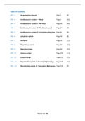
Table of contents
HFII - 1. Integumentary System Page 2 [8]
HFII - 2. Cardiovascular system I – Blood Page 1 [10]
HFII - 3. Cardiovascular system II - The heart Page 20 [14]
HFII - 4. Cardiovascular system III - The blood vessels Page 34 [7]
HFII - 5. Cardiovascular system IV - Circulatory physiology Page 41 [9]
HFII - 6. Lymphatic system Page 50 [8]
HFII - 7. Immunity Page 58 [12]
HFII - 8. Respiratory system Page 70 [20]
HFII - 9. Digestive system Page 90 [15]
HFII - 10. Urinary system Page 105 [11]
HFII - 11. Endocrinology Page 116 [12]
HFII – 12. Reproductive system I - Anatomy & physiology Page 128 [15]
HFII – 13. Reproductive system II – Conception & pregnancy Page 143 [9]
Pagina 1 van 188
, HFII - 1. Integumentary System
Integumentary system = organ system that protects body from damage:
• Mainly skin
• Accessory structures
o Cutaneous glands
o Sensory receptors
o Hair
o Nails
Some facts:
• Largest organ system
• Easy access for study and disease
• 6-8% of total body weight: 4.5-5.0 kg
• Covers 2m2
• Average thickness 1-2mm: thinnest (0.5mm) at eyelids and thickest (4.0mm) at heels
• Colour variety due to 3 pigments: melanin, haemoglobin, carotene ➔ determining factor is
melanin + balance pheomelanin (yellow to red) and eumelanin (brown to black).
o Permanent artificial colouring is possible when ink is deposited in the dermis
Main functions:
• External protection
• Blood storage reservoir
• Temperature regulation
• Cutaneous sensory detection
• Excretion and absorption
• Synthesis of vitamin D
Ancillary functions:
• Maintenance of structural integrity
• UV protection
• Non-verbal communication
Skin structure
The skin consists of:
Structure Tissue Function
Epidermis Avascular Keratinised stratified Protective barrier
Superficial squamous epithelium Water regulation
Thin (dead cells)
Dermis Vascular Connective tissue (CT) Holding the skin accessory structures
Thicker
Hypodermis Subcutaneous Areolar CT Stabilizes skin position: anchors dermis to fascia
Adipose tissue Contains blood supply to skin
Thermal insulation
Cushions underlying organs
Epidermis
The epidermis is arranged over 5 layers from out- to inside:
1) Stratum corneum
o Multiple layers (25-50 layers) of dead, flat keratinocytes
o Continuously shed and replaced from lower layers
o Form a hydrophobic protective layer
Pagina 2 van 188
, 2) Stratum lucidum
o Flattened keratinocytes (5-8 layers)
o Only present on thick skin ➔ palmar and plantar surfaces
o Thickened membranes to provide cushioning
3) Stratum granulosum
o Flattened keratinocytes (3-5 layers)
o Undergoing apoptosis as they move further from the blood stream
4) Stratum spinosum
o Keratinocytes gradually becoming flattened (8-10 layers)
o Keratin arranged into coarse filaments which bind cells together = tonofilaments
o This provides a strength and flexibility to resist tension
5) Stratum basale (germinal layer)
o Deepest layer of columnar keratinocytes and some stem cells attached to dermis
o Rapidly dividing cells gradually forming the upper layer cells
The epidermis has 4 cell types:
• Keratinocytes = deepest cells, arranged in layers and c90% of epidermal cell content
o Produce keratin through fibrous protein ➔ protection from microbial, thermal and
chemical damage
o Secrete lamellar granules that release a water repellent sealant ➔ maintains
osmotic integrity and regulates fluid movement
• Melanocytes = c8% of epidermal cell content
o Synthesise melanin ➔ contributes to skin colour and UV protection
o Long, slender projections extend between the keratinocytes for transfer of melanin
granules
• Merkel cells = tactile epithelial cells in low quantities
o Associated with sensory nerve endings in deep epidermis layers, forming Merkel
(tactile) discs ➔ detect touch sensations
• Langerhans cells = macrophage-like dendritic cells
o Participate in immune response
o Assist immune cells in protection
Pagina 3 van 188
,Dermis
The second dermis layer provides mechanical strength, flexibility, and protection for underlying
tissues due to collagen, elastic, and reticular fibres.
• Cell types: fibroblasts, macrophages, mast cells, white blood cells (WBCs)
• Highly vascular
• Variety of sensory receptors that provide information about the external environment
• Made up of a papillary and reticular layer
Papillary layer:
• Thinner component layer
• Composed of areolar CT
• Induces dermal papillae which contain capillary loops
o Some dermal papillae contain specialist touch receptors (Meissner corpuscles) and
free nerve endings
o Increase dermal surface area
• Supports and nourishes the epidermis
Reticular layer:
• Larger and thicker component layer
• Deeper layer representing 80% of dermis thickness
• Network of collagen and reticular fibres
o Network contains blood vessels, nerves, hair follicles, sweat glands, sebaceous
glands
o Also specialist deep touch receptors = Pacinian corpuscles and Ruffini endings
Hypodermis (subcutaneous layer)
The hypodermis is not strictly part of the true skin.
• Composed of loose CT ➔ adipose tissue (majority) and areolar CT (minority)
• Areolar CT is loosely attached to dermis and muscle ➔ stabilizes skin position
• The fat cells provide thermal insulation and cushions underlying organs
o They also cause dimpling of the skin (e.g. women’s hips, thighs) = cellulite
• Vascular ➔ target for subcutaneous injections because it absorbs drugs quickly
Pagina 4 van 188
,Skin thickness
Skin is almost uniform across the body, but there are some local variations due to:
• Epidermal thickness
• Keratinisation
• Distribution of glands
• Vascularity and nerve innervation
Thick skin is only found in areas of abrasion and pressure ➔ fingertips, palms, feet soles. It has an
extra epidermal layer (stratum lucidum) compared to thin skin.
Thin skin Thick skin
Distribution All body parts except palms and Palms, soles, palmar surfaces of
palmar surfaces of fingers digits
Epidermal thickness 0.10-0.15 mm 0.6-4.0 mm
Epidermal strata (layers) Thin strata spinosum and corneum All 5 strata
Absent stratum lucidum Thicker stratum spinosum and
grossly thicker stratum corneum
Epidermal ridges Lacking due to poorly developed Present due to numerous well-
dermal papillae organised dermal papillae
Hair follicles / Arrector pili Present Absent
Sebaceous glands Present Absent
Sudoriferous glands Few Numerous
Sensory receptors Sparse distribution Dense distribution
Arrector pili = small muscles attached to hair follicles
Sebaceous gland = microscopic exocrine gland in skin that secretes into a hair follicle to
lubricate hair and skin
Sudoriferous gland = sweat gland, produces sweat
Accessory structures
Hair, nails, cutaneous glands, and sensory receptors are accessory structures derived from the
epidermis but extend into the dermis.
Pagina 5 van 188
, Hair
Structure:
• Filamentous strands of dead keratinized cells produced by hair follicles
• Contains hard keratin ➔ tougher and more durable than soft keratin of the skin
• Consists of a root (embedded in the skin) and a shaft (projects above skin’s surface)
o Shaft has three concentric layers:
▪ Medulla = central core
▪ Cortex = surrounds medulla
▪ Cuticle = outermost layer
• Pigmented by melanocytes at the base of the hair
The hair root sheath extends from the epidermal surface into the dermis to form a hair bulb in the
follicle. The hair follicle consists of:
• Papilla = nipple-shaped indentation with blood vessels and nerves
• Matrix = germinal layer of actively dividing cells right above the papilla
• Root hair plexus = a knot of sensory nerve endings wrapping a hair bulb
o Bending of hair stimulates these nerves ➔ hairs are sensitive touch receptors
• Arrector pili muscle = bundle of smooth muscle contracts to make hair stand erect
Function of hair:
• Helps to maintain warmth
• Touch sensitivity
• Protection of scalp against physical trauma, heat loss, and sunlight
In adults hair is most distributed across the scalp, eyebrows, arm pits and external genitalia. No hair
grows on palms, soles, lips and nipples.
Pagina 6 van 188
HFII - 1. Integumentary System Page 2 [8]
HFII - 2. Cardiovascular system I – Blood Page 1 [10]
HFII - 3. Cardiovascular system II - The heart Page 20 [14]
HFII - 4. Cardiovascular system III - The blood vessels Page 34 [7]
HFII - 5. Cardiovascular system IV - Circulatory physiology Page 41 [9]
HFII - 6. Lymphatic system Page 50 [8]
HFII - 7. Immunity Page 58 [12]
HFII - 8. Respiratory system Page 70 [20]
HFII - 9. Digestive system Page 90 [15]
HFII - 10. Urinary system Page 105 [11]
HFII - 11. Endocrinology Page 116 [12]
HFII – 12. Reproductive system I - Anatomy & physiology Page 128 [15]
HFII – 13. Reproductive system II – Conception & pregnancy Page 143 [9]
Pagina 1 van 188
, HFII - 1. Integumentary System
Integumentary system = organ system that protects body from damage:
• Mainly skin
• Accessory structures
o Cutaneous glands
o Sensory receptors
o Hair
o Nails
Some facts:
• Largest organ system
• Easy access for study and disease
• 6-8% of total body weight: 4.5-5.0 kg
• Covers 2m2
• Average thickness 1-2mm: thinnest (0.5mm) at eyelids and thickest (4.0mm) at heels
• Colour variety due to 3 pigments: melanin, haemoglobin, carotene ➔ determining factor is
melanin + balance pheomelanin (yellow to red) and eumelanin (brown to black).
o Permanent artificial colouring is possible when ink is deposited in the dermis
Main functions:
• External protection
• Blood storage reservoir
• Temperature regulation
• Cutaneous sensory detection
• Excretion and absorption
• Synthesis of vitamin D
Ancillary functions:
• Maintenance of structural integrity
• UV protection
• Non-verbal communication
Skin structure
The skin consists of:
Structure Tissue Function
Epidermis Avascular Keratinised stratified Protective barrier
Superficial squamous epithelium Water regulation
Thin (dead cells)
Dermis Vascular Connective tissue (CT) Holding the skin accessory structures
Thicker
Hypodermis Subcutaneous Areolar CT Stabilizes skin position: anchors dermis to fascia
Adipose tissue Contains blood supply to skin
Thermal insulation
Cushions underlying organs
Epidermis
The epidermis is arranged over 5 layers from out- to inside:
1) Stratum corneum
o Multiple layers (25-50 layers) of dead, flat keratinocytes
o Continuously shed and replaced from lower layers
o Form a hydrophobic protective layer
Pagina 2 van 188
, 2) Stratum lucidum
o Flattened keratinocytes (5-8 layers)
o Only present on thick skin ➔ palmar and plantar surfaces
o Thickened membranes to provide cushioning
3) Stratum granulosum
o Flattened keratinocytes (3-5 layers)
o Undergoing apoptosis as they move further from the blood stream
4) Stratum spinosum
o Keratinocytes gradually becoming flattened (8-10 layers)
o Keratin arranged into coarse filaments which bind cells together = tonofilaments
o This provides a strength and flexibility to resist tension
5) Stratum basale (germinal layer)
o Deepest layer of columnar keratinocytes and some stem cells attached to dermis
o Rapidly dividing cells gradually forming the upper layer cells
The epidermis has 4 cell types:
• Keratinocytes = deepest cells, arranged in layers and c90% of epidermal cell content
o Produce keratin through fibrous protein ➔ protection from microbial, thermal and
chemical damage
o Secrete lamellar granules that release a water repellent sealant ➔ maintains
osmotic integrity and regulates fluid movement
• Melanocytes = c8% of epidermal cell content
o Synthesise melanin ➔ contributes to skin colour and UV protection
o Long, slender projections extend between the keratinocytes for transfer of melanin
granules
• Merkel cells = tactile epithelial cells in low quantities
o Associated with sensory nerve endings in deep epidermis layers, forming Merkel
(tactile) discs ➔ detect touch sensations
• Langerhans cells = macrophage-like dendritic cells
o Participate in immune response
o Assist immune cells in protection
Pagina 3 van 188
,Dermis
The second dermis layer provides mechanical strength, flexibility, and protection for underlying
tissues due to collagen, elastic, and reticular fibres.
• Cell types: fibroblasts, macrophages, mast cells, white blood cells (WBCs)
• Highly vascular
• Variety of sensory receptors that provide information about the external environment
• Made up of a papillary and reticular layer
Papillary layer:
• Thinner component layer
• Composed of areolar CT
• Induces dermal papillae which contain capillary loops
o Some dermal papillae contain specialist touch receptors (Meissner corpuscles) and
free nerve endings
o Increase dermal surface area
• Supports and nourishes the epidermis
Reticular layer:
• Larger and thicker component layer
• Deeper layer representing 80% of dermis thickness
• Network of collagen and reticular fibres
o Network contains blood vessels, nerves, hair follicles, sweat glands, sebaceous
glands
o Also specialist deep touch receptors = Pacinian corpuscles and Ruffini endings
Hypodermis (subcutaneous layer)
The hypodermis is not strictly part of the true skin.
• Composed of loose CT ➔ adipose tissue (majority) and areolar CT (minority)
• Areolar CT is loosely attached to dermis and muscle ➔ stabilizes skin position
• The fat cells provide thermal insulation and cushions underlying organs
o They also cause dimpling of the skin (e.g. women’s hips, thighs) = cellulite
• Vascular ➔ target for subcutaneous injections because it absorbs drugs quickly
Pagina 4 van 188
,Skin thickness
Skin is almost uniform across the body, but there are some local variations due to:
• Epidermal thickness
• Keratinisation
• Distribution of glands
• Vascularity and nerve innervation
Thick skin is only found in areas of abrasion and pressure ➔ fingertips, palms, feet soles. It has an
extra epidermal layer (stratum lucidum) compared to thin skin.
Thin skin Thick skin
Distribution All body parts except palms and Palms, soles, palmar surfaces of
palmar surfaces of fingers digits
Epidermal thickness 0.10-0.15 mm 0.6-4.0 mm
Epidermal strata (layers) Thin strata spinosum and corneum All 5 strata
Absent stratum lucidum Thicker stratum spinosum and
grossly thicker stratum corneum
Epidermal ridges Lacking due to poorly developed Present due to numerous well-
dermal papillae organised dermal papillae
Hair follicles / Arrector pili Present Absent
Sebaceous glands Present Absent
Sudoriferous glands Few Numerous
Sensory receptors Sparse distribution Dense distribution
Arrector pili = small muscles attached to hair follicles
Sebaceous gland = microscopic exocrine gland in skin that secretes into a hair follicle to
lubricate hair and skin
Sudoriferous gland = sweat gland, produces sweat
Accessory structures
Hair, nails, cutaneous glands, and sensory receptors are accessory structures derived from the
epidermis but extend into the dermis.
Pagina 5 van 188
, Hair
Structure:
• Filamentous strands of dead keratinized cells produced by hair follicles
• Contains hard keratin ➔ tougher and more durable than soft keratin of the skin
• Consists of a root (embedded in the skin) and a shaft (projects above skin’s surface)
o Shaft has three concentric layers:
▪ Medulla = central core
▪ Cortex = surrounds medulla
▪ Cuticle = outermost layer
• Pigmented by melanocytes at the base of the hair
The hair root sheath extends from the epidermal surface into the dermis to form a hair bulb in the
follicle. The hair follicle consists of:
• Papilla = nipple-shaped indentation with blood vessels and nerves
• Matrix = germinal layer of actively dividing cells right above the papilla
• Root hair plexus = a knot of sensory nerve endings wrapping a hair bulb
o Bending of hair stimulates these nerves ➔ hairs are sensitive touch receptors
• Arrector pili muscle = bundle of smooth muscle contracts to make hair stand erect
Function of hair:
• Helps to maintain warmth
• Touch sensitivity
• Protection of scalp against physical trauma, heat loss, and sunlight
In adults hair is most distributed across the scalp, eyebrows, arm pits and external genitalia. No hair
grows on palms, soles, lips and nipples.
Pagina 6 van 188



