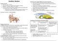
Auditory System How the ear can recognize different sound wave frequencies
● Outer ear channels sound waves up into the tympanic membrane
Anatomy of the Ear ● Sound waves will make the tympanic membrane vibrate the ossicles (to
● Outer ear : pinna, ear canal overcome impedance)
● Outer and middle ear is separated by the tympanic membrane ● Ossicles turn air vibrations into mechanical vibrations
● Middle ear : 3 ossicles (malleus, incus, stapes) ● Vibrations then get transmitted through the oval window, directed into the
● Inner ear : behind the oval window, containing membranous fluid filled space scala vestibuli and go up through the cochlea
in the temporal bone, cochlea, cochlear duct ● Vibrations are directed into the scala vestibuli
○ Bony labyrinth in temporal bone filled with perilymph (high Na, low K) ● Basilar membrane will start vibrating
○ Membranous labyrinth filled with endolymph (high K, low Na) ● Side by side tilting of stereocilia that are embedded into the basilar
membrane
Endolymph is produced in the stria vascularis
Transduction of sound waves into electrical signals
At Rest When tip links are stretched When tip links tilt in the
(strong vibrations) opposite direction (weaker
vibrations or stopped)
Anatomy of the inner ear
a. Cochlea (auditory part of the inner ear) : base (receptive to high frequency) spirals Slightly depolarized More Na channels open = Close Na channels =
up to the apex (receptive to low frequency) and spirals up 2.5 times (-40mV) due to open increased Na influx hyperpolarize
● Function : tell the different sound frequencies Na channels at the
top of hair cells Excessive positive charge
● Cochlear branch of the vestibulocochlear (VIII) nerve runs through middle of
in endolymph drives Na
cochlea and innervates the membranous labyrinth Hair cell leaks influx = more depolarization
● 3 chambers of the cochlea glutamate into
○ Scala Vestibuli (upper chamber) : filled with perilymph afferent nerve = firing More glutamate release =
○ Scala Tympani (lower chamber) : filled with perilymph AP increased AP firing
○ Cochlear duct (internal chamber): filled with endolymph
● Auditory hair cells (1 layer of inner hair cells , several layers of outer hair ● Louder sounds produce larger vibration → bigger receptor potentials = more
cells) are in the spiral organ NT release, creating more APs
b. Tectorial membrane : gelatinous membrane that lies over the top of hair cells ● Different frequencies (pitch) activate different hair cells
important for opening and closing hair cells
● Outer hair cells are embedded inside the tectorial membrane and link it to the
spiral organ and basilar membrane
● It also links the stereocilia together with glycoproteins (for depolarization)
● Outer ear channels sound waves up into the tympanic membrane
Anatomy of the Ear ● Sound waves will make the tympanic membrane vibrate the ossicles (to
● Outer ear : pinna, ear canal overcome impedance)
● Outer and middle ear is separated by the tympanic membrane ● Ossicles turn air vibrations into mechanical vibrations
● Middle ear : 3 ossicles (malleus, incus, stapes) ● Vibrations then get transmitted through the oval window, directed into the
● Inner ear : behind the oval window, containing membranous fluid filled space scala vestibuli and go up through the cochlea
in the temporal bone, cochlea, cochlear duct ● Vibrations are directed into the scala vestibuli
○ Bony labyrinth in temporal bone filled with perilymph (high Na, low K) ● Basilar membrane will start vibrating
○ Membranous labyrinth filled with endolymph (high K, low Na) ● Side by side tilting of stereocilia that are embedded into the basilar
membrane
Endolymph is produced in the stria vascularis
Transduction of sound waves into electrical signals
At Rest When tip links are stretched When tip links tilt in the
(strong vibrations) opposite direction (weaker
vibrations or stopped)
Anatomy of the inner ear
a. Cochlea (auditory part of the inner ear) : base (receptive to high frequency) spirals Slightly depolarized More Na channels open = Close Na channels =
up to the apex (receptive to low frequency) and spirals up 2.5 times (-40mV) due to open increased Na influx hyperpolarize
● Function : tell the different sound frequencies Na channels at the
top of hair cells Excessive positive charge
● Cochlear branch of the vestibulocochlear (VIII) nerve runs through middle of
in endolymph drives Na
cochlea and innervates the membranous labyrinth Hair cell leaks influx = more depolarization
● 3 chambers of the cochlea glutamate into
○ Scala Vestibuli (upper chamber) : filled with perilymph afferent nerve = firing More glutamate release =
○ Scala Tympani (lower chamber) : filled with perilymph AP increased AP firing
○ Cochlear duct (internal chamber): filled with endolymph
● Auditory hair cells (1 layer of inner hair cells , several layers of outer hair ● Louder sounds produce larger vibration → bigger receptor potentials = more
cells) are in the spiral organ NT release, creating more APs
b. Tectorial membrane : gelatinous membrane that lies over the top of hair cells ● Different frequencies (pitch) activate different hair cells
important for opening and closing hair cells
● Outer hair cells are embedded inside the tectorial membrane and link it to the
spiral organ and basilar membrane
● It also links the stereocilia together with glycoproteins (for depolarization)