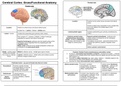
Cerebral Cortex: Gross/Functional Anatomy Parietal lobe
Frontal Lobe
Location Posterior to the central sulcus and above the lateral
sulcus
Parieto-occipital sulcus separates it from occipital lobe,
Location Anterior to central sulcus and above lateral sulcus but is only visible in the medial aspect of the
hemisphere
Divided into 1. Lateral 2. Medial 3. Orbital parts
Lateral parietal region Contains postcentral gyrus (primary somatosensory
Lateral : 2 parts Contains the precentral gyrus (primary motor cortex) cortex)
Rest consists of 1. Superior 2. Middle 3. Inferior frontal gyri, Posterior parietal cortex Superior parietal cortex/lobule : continues onto
separated by superior and inferior frontal sulci (somatosensory association medial surface of hemisphere as the rectangular
cortex) : divided by intraparietal shaped precuneus
Superior frontal gyrus continues onto the medial surface of the sulcus (deep cleft oriented at right *boundaries of precuneus : paracentral lobule (front),
hemisphere and is separated from the cingulate gyrus with angles to central sulcus) occipital lobe (behind), posterior cingulate cortex
cingulate sulcus. (below)
Motor cortex continues to the medial hemispheric surface and
contributes to the paracentral lobule Inferior parietal cortex/lobule)
Orbital : overlies orbital Medial to olfactory sulcus : straight gyrus (gyrus rectus) Posterior Parietal Cortex (somatosensory association cortex)
cavities and is divided ● Involved in visuospatial representation of objects in extrapersonal space → receives
into 2 areas by olfactory Lateral to olfactory sulcus : orbital cortex and contains a poorly projections from occipital lobes which identify where the objects are and how they move
sulcus defined H-shaped orbital sulcus with respect to us
● Important for object interaction (eg. reach, grasp, rotation) → identifies objects in the
environment by using visual attention and voluntary gaze
● Important to initiate semi-automatic motor sequences (praxes)
Prefrontal Cortex : any part of frontal lobe that isn’t motor
Inferior parietal cortex Involved in symbolic Lesions cause Gerstmann
Dorsolateral Controls cognitive function representation, concept of Syndrome → difficulty with
(thinking/intellect) and frontal numbers (math and physics simple math, left/right
eye fields (attention, gaze, ability), reading/writing confusion, difficulty naming
voluntary eye movement) individual fingers
Orbitofrontal Controls behavior, Medial parietal cortex Involved in day dreaming, quiet Low activity in aphantasia
personality, and social (precuneus) contemplation, meditation, recall (inability to visualize)
Medial conduct (inhibition, restraint, of episodic memory, and
Prefrontal and tact) visualization of real/imaginary
objects (ie. mind’s eye)
, Praxes : motor programme (eg. tying shoelaces) stored in the premotor cortex by selected by the WHAT (temporal) vs WHERE (parietal) Pathways
posterior parietal lobule
Damage to the posterior parietal cortex → deficits in ordinary daily tasks (eg. using fork/knife, Agnosia (failure to recognize objects) has a
getting dressed) but are anosognosic (unaware of neurological problem)and problem in which pathway? The WHAT (temporal)
● Stroke : associated with attentional problems (eg. read ‘peanut’ as ‘nut’)
● Classical neglect syndrome caused by right parietal lesion : only eat left side of the plate
Occipital Lobe : entirely associated with vision
Temporal Lobe
Location Projects downwards+forwards, below the
lateral sulcus.
Posteriorly bounded by preoccipital notch
Lateral region is divided into 1. Superior 2.
Middle 3. Inferior temporal gyri by superior
and inferior temporal sulci
Location Posterior to the parieto-occipital sulcus and preoccipital
notch Superior 1+ transverse temporal gyri are identified
Most exposed surface of occipital lobe consists of visual surface : posteriorly and medially
association cortex flat and
hidden They have roles in :
1. Medial 2. Lateral 3. Inferior surfaces inside lateral 1. Wernicke’s Area (speech recognition)
sulcus 2. Object recognition/categorization
Medial occipital lobe : divided Cuneus 3. Comprehension
into 2 areas by calcarine
sulcus Lingual gyrus
Broca’s Area Wernicke’s Area
Primary visual cortex Lies in the crease of the sulcus
Location Inferior frontal gyrus In posterior ⅓ of superior
*usually on the (triangular/opercular parts) temporal gyrus (part of auditory
Motor Landmarks : inverted capital omega Sensory Landmarks left side association cortex
corresponding to part of motor cortex 1. Somatosensory : behind central gyrus
controlling the contralateral hand (hand 2. Auditory : anterior transverse temporal Connected by arcuate fasciculus which begins in the frontal lobe,
knob) gyri on superior surface of temporal lobe passes posteriorly, and sweeps down into temporal lobe
3. Visual : crease of calcarine sulcus
What is it? Motor language area → involved Sensory language area →
in production of speech/language, involved in recognition of speech
ensuring utterances follow sounds and understanding of
grammar/syntax rules to the spoken language
listener understands
Adjacent to the Wernicke’s area (in inferior parietal lobe) is involved in understanding written
language
Frontal Lobe
Location Posterior to the central sulcus and above the lateral
sulcus
Parieto-occipital sulcus separates it from occipital lobe,
Location Anterior to central sulcus and above lateral sulcus but is only visible in the medial aspect of the
hemisphere
Divided into 1. Lateral 2. Medial 3. Orbital parts
Lateral parietal region Contains postcentral gyrus (primary somatosensory
Lateral : 2 parts Contains the precentral gyrus (primary motor cortex) cortex)
Rest consists of 1. Superior 2. Middle 3. Inferior frontal gyri, Posterior parietal cortex Superior parietal cortex/lobule : continues onto
separated by superior and inferior frontal sulci (somatosensory association medial surface of hemisphere as the rectangular
cortex) : divided by intraparietal shaped precuneus
Superior frontal gyrus continues onto the medial surface of the sulcus (deep cleft oriented at right *boundaries of precuneus : paracentral lobule (front),
hemisphere and is separated from the cingulate gyrus with angles to central sulcus) occipital lobe (behind), posterior cingulate cortex
cingulate sulcus. (below)
Motor cortex continues to the medial hemispheric surface and
contributes to the paracentral lobule Inferior parietal cortex/lobule)
Orbital : overlies orbital Medial to olfactory sulcus : straight gyrus (gyrus rectus) Posterior Parietal Cortex (somatosensory association cortex)
cavities and is divided ● Involved in visuospatial representation of objects in extrapersonal space → receives
into 2 areas by olfactory Lateral to olfactory sulcus : orbital cortex and contains a poorly projections from occipital lobes which identify where the objects are and how they move
sulcus defined H-shaped orbital sulcus with respect to us
● Important for object interaction (eg. reach, grasp, rotation) → identifies objects in the
environment by using visual attention and voluntary gaze
● Important to initiate semi-automatic motor sequences (praxes)
Prefrontal Cortex : any part of frontal lobe that isn’t motor
Inferior parietal cortex Involved in symbolic Lesions cause Gerstmann
Dorsolateral Controls cognitive function representation, concept of Syndrome → difficulty with
(thinking/intellect) and frontal numbers (math and physics simple math, left/right
eye fields (attention, gaze, ability), reading/writing confusion, difficulty naming
voluntary eye movement) individual fingers
Orbitofrontal Controls behavior, Medial parietal cortex Involved in day dreaming, quiet Low activity in aphantasia
personality, and social (precuneus) contemplation, meditation, recall (inability to visualize)
Medial conduct (inhibition, restraint, of episodic memory, and
Prefrontal and tact) visualization of real/imaginary
objects (ie. mind’s eye)
, Praxes : motor programme (eg. tying shoelaces) stored in the premotor cortex by selected by the WHAT (temporal) vs WHERE (parietal) Pathways
posterior parietal lobule
Damage to the posterior parietal cortex → deficits in ordinary daily tasks (eg. using fork/knife, Agnosia (failure to recognize objects) has a
getting dressed) but are anosognosic (unaware of neurological problem)and problem in which pathway? The WHAT (temporal)
● Stroke : associated with attentional problems (eg. read ‘peanut’ as ‘nut’)
● Classical neglect syndrome caused by right parietal lesion : only eat left side of the plate
Occipital Lobe : entirely associated with vision
Temporal Lobe
Location Projects downwards+forwards, below the
lateral sulcus.
Posteriorly bounded by preoccipital notch
Lateral region is divided into 1. Superior 2.
Middle 3. Inferior temporal gyri by superior
and inferior temporal sulci
Location Posterior to the parieto-occipital sulcus and preoccipital
notch Superior 1+ transverse temporal gyri are identified
Most exposed surface of occipital lobe consists of visual surface : posteriorly and medially
association cortex flat and
hidden They have roles in :
1. Medial 2. Lateral 3. Inferior surfaces inside lateral 1. Wernicke’s Area (speech recognition)
sulcus 2. Object recognition/categorization
Medial occipital lobe : divided Cuneus 3. Comprehension
into 2 areas by calcarine
sulcus Lingual gyrus
Broca’s Area Wernicke’s Area
Primary visual cortex Lies in the crease of the sulcus
Location Inferior frontal gyrus In posterior ⅓ of superior
*usually on the (triangular/opercular parts) temporal gyrus (part of auditory
Motor Landmarks : inverted capital omega Sensory Landmarks left side association cortex
corresponding to part of motor cortex 1. Somatosensory : behind central gyrus
controlling the contralateral hand (hand 2. Auditory : anterior transverse temporal Connected by arcuate fasciculus which begins in the frontal lobe,
knob) gyri on superior surface of temporal lobe passes posteriorly, and sweeps down into temporal lobe
3. Visual : crease of calcarine sulcus
What is it? Motor language area → involved Sensory language area →
in production of speech/language, involved in recognition of speech
ensuring utterances follow sounds and understanding of
grammar/syntax rules to the spoken language
listener understands
Adjacent to the Wernicke’s area (in inferior parietal lobe) is involved in understanding written
language