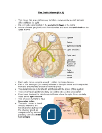
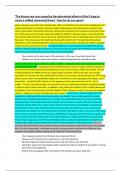
The Optic Nerve (CN II)
This nerve has a special sensory function, carrying only special somatic
afferent fibres for sight
It’s cell bodies are located in the ganglionic layer of the retina
Axons of these ganglionic cells form bundles and leave the optic bulb as the
optic nerve
Each optic nerve contains around 1 million myelinated axons
Part of the meninges (pia mater) adheres to the optic nerve and is separated
from the arachnoid by the subarachnoid space
The dura forms an outer sheath and fuses with the sclera of the eyeball
The nerve travels posteromedially to exit the orbit via the optic canal
From here it enters the middle cranial fossa where the optic fibres partially
cross at the optic chiasm
This enables humans to have
binocular vision
The optic chiasm is found
directly inferior to the
hypothalamus but superior to
the pituitary gland, so
compression by an enlarging
pituitary can cause bitemporal
hemianopia