Lecture 1: Serum Albumin
Human Serum Albumin
- major protein present in blood plasma
>50% of protein present
- two major functions:
1) transports smaller, mostly hydrophobic molecules
2) major contributor of osmotic swelling pressure of blood plasma
- large amount of serum albumin excludes water resulting in osmotic swelling
- osmotic swelling: positive pressure in bloodstream
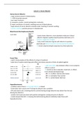
Blood Serum Electrophoresis Results
- clearly shows albumin is most abundant molecule in blood
- albumin migrates rapidly towards positive electrode as it’s
surface is negatively charged
- many small molecules migrate with albumin to positive
electrode and they’re bound to and transported by albumin
remain attached despite separation by electrophoresis
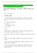
Forest Plots
- used in meta-analysis of the effects of a drug or treatment
- takes lots of studies examining same effect and shows outcome of them all added together
- odds ratio (OR): ratio between effect in test sample &
effect in control
control can be no placebo, treatment, or effects
from already developed drugs
- OR = 1: no effect, test & control same
- OR > 1: test better than control
- OR < 1: control better than test
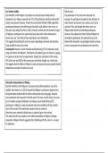
Hypoalbuminemia
- condition where there’s not enough albumin in blood
- forest plots were used to see if having low albumin was a problem
- OR came back to be 2 showing that the control (having enough albumin) was better than the test
(having low albumin)
- another forest plot was done with patients being given varying amounts of albumin
- results showed that OR > 1 showing that administering albumin definitely helps those with
hypoalbuminemia
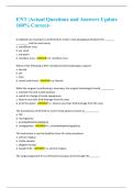
,α-Domain Structures
- protein structures entirely composed of α-helices
- α-helices aren’t very stable in solution as they tend to unwind so they interact with other
structures to stabilise themselves
- α-helices are therefore packed pairwise in protein with hydrophobic residues pointing towards the
molecules core
- Rop – small protein that binds with RNA
- hydrophobic residues (green) are all pointed
to the core
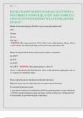
Structure of Serum Albumin
- HSA Mr = 66,500 kilodaltons
- pI = 5.67 meaning it’s quite negatively charged at a physiological pH
- composed almost entirely of α-helices
67% α-helical
- consists of single polypeptide chain containing 585 amino acids with 17 intra-chain disulphide
bonds
disulphide bonds formed between two cysteine residues meaning there are 34 cysteine residues
making the bonds
- disulphide bonds pull molecule into series of large & small loops
- three similar domains in serum albumin but amino acid sequence repeats even from one individual
loop to another
- 2D early structure of albumin
- loops in all three domains are L, S, L
longer loops have longer α-helices and shorter loops have shorter ones
Evolution
- original gene duplicated & mutated until the HSA LSL-
LSL-LSL structure was formed
- through evolution, HSA must’ve had a continuous
important function to be kept in the genome
,Cysteines
- amino acid sequence of albumin has unusually high % of cysteine with 35 out of 585
35 out of 35 form disulphide bonds
- cysteine very reactive amino acid due to lone pair on S that really want to form covalent bonds
- in plasma, 30% of free thiol group on Cys-34 is oxidised
Albumin as Transporter
- albumin contains many binding sites esp. for long chain fatty acids, smaller heterocyclic or aromatic
COOHs & metal
- the SH of Cys-34 can bind Cd, Au, Hg & Ag
- the N-terminal binds Cu(II), Ni(II), Ca & Zn(II)
- at the beginning of loop 1, the first three amino acid residues are very effective at
binding to Cu
- lone pair on N is donated to Cu forming a complex which is how most copper is
transported around body
Drug Transport
- as albumin has good binding properties, it can transport many hormones & drugs around the body
e.g. aspirin, penicillin etc
- pharmacologically important as drugs compete for binding sites
- if drug molecules don’t bind to HSA, they’ll be excreted by the kidneys
, Human Serum Albumin – 3D Structure
- molecule adopts heart shape with 3 domains
- domain I in red
- domain II in green
- domain III in blue
- even though I & III are separated from each other, they
pack up against each other forming a packed, globular shape
- this particular structure has no ligands bound
Fatty Acid Binding
- albumin structure with myristic acid bound
- four molecules of this fatty acid can bind to hydrophobic
pockets within loops of the α-helices
Confirmation Change Upon Binding
- the shape of the HSA changes upon the binding of ligands
- albumin can change itself to accommodate ligands binding
or the ligands change the shape of the albumin
- may enable it to hold on tighter to the ligand or to
encourage another ligand to bind or may have a role in
telling the HSA where to transport the molecules
Ligands Binding
- fatty acid in held in empty region between
3-4 α-helices
- spaces created by hydrophobic residues of
HSA
Human Serum Albumin
- major protein present in blood plasma
>50% of protein present
- two major functions:
1) transports smaller, mostly hydrophobic molecules
2) major contributor of osmotic swelling pressure of blood plasma
- large amount of serum albumin excludes water resulting in osmotic swelling
- osmotic swelling: positive pressure in bloodstream
Blood Serum Electrophoresis Results
- clearly shows albumin is most abundant molecule in blood
- albumin migrates rapidly towards positive electrode as it’s
surface is negatively charged
- many small molecules migrate with albumin to positive
electrode and they’re bound to and transported by albumin
remain attached despite separation by electrophoresis
Forest Plots
- used in meta-analysis of the effects of a drug or treatment
- takes lots of studies examining same effect and shows outcome of them all added together
- odds ratio (OR): ratio between effect in test sample &
effect in control
control can be no placebo, treatment, or effects
from already developed drugs
- OR = 1: no effect, test & control same
- OR > 1: test better than control
- OR < 1: control better than test
Hypoalbuminemia
- condition where there’s not enough albumin in blood
- forest plots were used to see if having low albumin was a problem
- OR came back to be 2 showing that the control (having enough albumin) was better than the test
(having low albumin)
- another forest plot was done with patients being given varying amounts of albumin
- results showed that OR > 1 showing that administering albumin definitely helps those with
hypoalbuminemia
,α-Domain Structures
- protein structures entirely composed of α-helices
- α-helices aren’t very stable in solution as they tend to unwind so they interact with other
structures to stabilise themselves
- α-helices are therefore packed pairwise in protein with hydrophobic residues pointing towards the
molecules core
- Rop – small protein that binds with RNA
- hydrophobic residues (green) are all pointed
to the core
Structure of Serum Albumin
- HSA Mr = 66,500 kilodaltons
- pI = 5.67 meaning it’s quite negatively charged at a physiological pH
- composed almost entirely of α-helices
67% α-helical
- consists of single polypeptide chain containing 585 amino acids with 17 intra-chain disulphide
bonds
disulphide bonds formed between two cysteine residues meaning there are 34 cysteine residues
making the bonds
- disulphide bonds pull molecule into series of large & small loops
- three similar domains in serum albumin but amino acid sequence repeats even from one individual
loop to another
- 2D early structure of albumin
- loops in all three domains are L, S, L
longer loops have longer α-helices and shorter loops have shorter ones
Evolution
- original gene duplicated & mutated until the HSA LSL-
LSL-LSL structure was formed
- through evolution, HSA must’ve had a continuous
important function to be kept in the genome
,Cysteines
- amino acid sequence of albumin has unusually high % of cysteine with 35 out of 585
35 out of 35 form disulphide bonds
- cysteine very reactive amino acid due to lone pair on S that really want to form covalent bonds
- in plasma, 30% of free thiol group on Cys-34 is oxidised
Albumin as Transporter
- albumin contains many binding sites esp. for long chain fatty acids, smaller heterocyclic or aromatic
COOHs & metal
- the SH of Cys-34 can bind Cd, Au, Hg & Ag
- the N-terminal binds Cu(II), Ni(II), Ca & Zn(II)
- at the beginning of loop 1, the first three amino acid residues are very effective at
binding to Cu
- lone pair on N is donated to Cu forming a complex which is how most copper is
transported around body
Drug Transport
- as albumin has good binding properties, it can transport many hormones & drugs around the body
e.g. aspirin, penicillin etc
- pharmacologically important as drugs compete for binding sites
- if drug molecules don’t bind to HSA, they’ll be excreted by the kidneys
, Human Serum Albumin – 3D Structure
- molecule adopts heart shape with 3 domains
- domain I in red
- domain II in green
- domain III in blue
- even though I & III are separated from each other, they
pack up against each other forming a packed, globular shape
- this particular structure has no ligands bound
Fatty Acid Binding
- albumin structure with myristic acid bound
- four molecules of this fatty acid can bind to hydrophobic
pockets within loops of the α-helices
Confirmation Change Upon Binding
- the shape of the HSA changes upon the binding of ligands
- albumin can change itself to accommodate ligands binding
or the ligands change the shape of the albumin
- may enable it to hold on tighter to the ligand or to
encourage another ligand to bind or may have a role in
telling the HSA where to transport the molecules
Ligands Binding
- fatty acid in held in empty region between
3-4 α-helices
- spaces created by hydrophobic residues of
HSA