
ANATOMY OF THE HEART AND MEDIASTINUM
Mediastinum is found within the thoracic cavity
Average rate of heart contractions is 75 per minute
= 108,000 times a day and over 39 million times a year
At least 75ml of blood is ejected per contraction
= 5.25L per min being pumped around 97,000 km of vessels in the body
The heart sits in the pericardial cavity (peri=top) created by the pericardial sac which is a
tough membrane that surrounds the heart.
The pericardial sac separates the heart from the rest of the structures in the mediastinum.
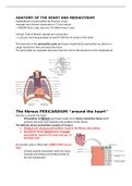
The fibrous PERICARDIUM ‘’around the heart’’
Directly surrounds the heart
- Pericardium is FIBROUS and tough made out of dense connective tissue which
protects the heart and maintains the position of the thorax.
The delicate serous pericardium consists of 2 layers:
1. Parietal layer of serous pericardium: fused to the fibrous pericardium
2. Epicardium (Inner visceral layer of serous)
pericardium: fused to the heart and part of
the heart wall
Pericardial cavity is filled with LUBRICATING serous
fluid
- Creates smooth movement when the heart
expands and relaxes preventing friction of
the two membranes
, BASE AND APEX
The superior surface of the heart is called the base and the inferior tip is called the apex.
The great veins and arteries are found and attached at
the base of the heart
- This is at the level of the third costal cartilage
(ribs)
Superior and inferior vena cava
Aorta
Pulmonary arteries and veins
Apex lies to the direct left of the sternum (long bone
between ribs) between the fourth and fifth ribs.
- It points into the left lung creating an
indentation on its medial surface
The average heart is about 12 x 8 x 6cm
250-300g in weight
The heart is made up of cardiac muscle meaning it increases in
cell size with response to exercise
HYPERTROPHIC CARDIOMYOPATHY:
The heart is abnormally enlarged with no exact cause.
CHAMBERS OF THE HEART:
Mediastinum is found within the thoracic cavity
Average rate of heart contractions is 75 per minute
= 108,000 times a day and over 39 million times a year
At least 75ml of blood is ejected per contraction
= 5.25L per min being pumped around 97,000 km of vessels in the body
The heart sits in the pericardial cavity (peri=top) created by the pericardial sac which is a
tough membrane that surrounds the heart.
The pericardial sac separates the heart from the rest of the structures in the mediastinum.
The fibrous PERICARDIUM ‘’around the heart’’
Directly surrounds the heart
- Pericardium is FIBROUS and tough made out of dense connective tissue which
protects the heart and maintains the position of the thorax.
The delicate serous pericardium consists of 2 layers:
1. Parietal layer of serous pericardium: fused to the fibrous pericardium
2. Epicardium (Inner visceral layer of serous)
pericardium: fused to the heart and part of
the heart wall
Pericardial cavity is filled with LUBRICATING serous
fluid
- Creates smooth movement when the heart
expands and relaxes preventing friction of
the two membranes
, BASE AND APEX
The superior surface of the heart is called the base and the inferior tip is called the apex.
The great veins and arteries are found and attached at
the base of the heart
- This is at the level of the third costal cartilage
(ribs)
Superior and inferior vena cava
Aorta
Pulmonary arteries and veins
Apex lies to the direct left of the sternum (long bone
between ribs) between the fourth and fifth ribs.
- It points into the left lung creating an
indentation on its medial surface
The average heart is about 12 x 8 x 6cm
250-300g in weight
The heart is made up of cardiac muscle meaning it increases in
cell size with response to exercise
HYPERTROPHIC CARDIOMYOPATHY:
The heart is abnormally enlarged with no exact cause.
CHAMBERS OF THE HEART:


