
Topic 1: Lifestyle, Health and Risk / Pearson Edexcel Biology A (Salters-Nuffield) / 9BN0
Mass transport & water
1.1-1.2 / circulation overcomes the limits of diffusion
Why do large animals need circulation?
Larger animal -> lower surface area to volume ratio -> longer diffusion distances. Diffusion
alone cannot supply distant, active cells fast enough.
Heart generates pressure -> blood moves by mass flow -> capillaries bring oxygen and
nutrients close to cells. Diffusion completes exchange over the final short distance; blood
carries CO2 and other wastes away.
Double circulation in mammals
Pulmonary circuit Right heart -> lungs -> left heart
Systemic circuit Left heart -> body tissues -> right heart
Blood passes through the heart twice per complete circuit. Separate circuits limit mixing
and allow higher pressure in the systemic circuit.
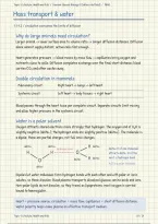
Water is a polar solvent
Oxygen attracts shared electrons more strongly than hydrogen. The oxygen end of H2O is
slightly negative (delta-); the hydrogen ends are slightly positive (delta+). The molecule is
a dipole: these are partial charges, not full ionic charges.
delta+ delta+
hydrogen bond
H delta+
delta+ H of one molecule
H H O attracts delta- O of the
O delta- H delta+ next: a hydrogen bond
H2O is a polar molecule
delta-
Dipoles let water molecules form hydrogen bonds with each other and with polar or ionic
solutes, so these dissolve: blood plasma transports dissolved glucose, amino acids and ions.
Non-polar lipids do not dissolve, so they travel as lipoproteins; most oxygen is carried
bound to haemoglobin.
Heart = pressure source; circulation = mass flow; capillaries = short diffusion distance.
Water polarity helps make plasma an effective transport medium.
Topic 1: Lifestyle, Health and Risk
,Topic 1: Lifestyle, Health and Risk / Pearson Edexcel Biology A (Salters-Nuffield) / 9BN0
Blood vessels
1.3 / structure -> function comparison
Arteries: away from the heart
High pressure Thick wall and collagen resist excessive stretching.
Elastic tissue Stretches in systole; recoils in diastole -> maintains pressure
and smooths flow.
Smooth muscle Alters lumen diameter. Arterioles control resistance and
blood distribution.
Veins: towards the heart
Low pressure Thin wall, less muscle and elastic tissue; large lumen -> low
resistance.
Valves + muscles Skeletal muscles compress veins; valves prevent backflow
towards tissues.
Capillaries: exchange with tissues
Single endothelial layer Short diffusion distance; permeability varies between tissues.
Small lumen + branches Blood stays close to the wall. Large total surface area and
cross-sectional area -> slower flow and more time for
exchange.
Arteries have a narrow lumen relative to wall thickness; veins often have a larger, less
regular lumen. Endothelium is smooth to reduce friction. Use several features because
tissue preparation can distort shape.
Direction defines artery vs. vein. Pulmonary artery: deoxygenated blood. Pulmonary
veins: oxygenated blood. Oxygen and glucose move towards tissues; wastes move into
blood.
Topic 1: Lifestyle, Health and Risk
, Topic 1: Lifestyle, Health and Risk / Pearson Edexcel Biology A (Salters-Nuffield) / 9BN0
Vessel structure: diagrams
1.3 / artery cross-section and capillary cutaway
Artery: thick wall around the lumen
outer layer: collagen
smooth muscle + elastic fibres
endothelium: inner lining
lumen containing blood
Collagen supports the wall; smooth muscle alters diameter; elastic tissue stretches and
recoils. Use the layer positions rather than memorising the illustration colours.
Capillary: one layer of endothelial cells
basement membrane (not a cell layer)
endothelial cell cell nucleus
A single endothelial layer gives a short diffusion distance. A narrow lumen keeps blood
close to the wall; many capillaries provide a large exchange surface area.
Topic 1: Lifestyle, Health and Risk
Mass transport & water
1.1-1.2 / circulation overcomes the limits of diffusion
Why do large animals need circulation?
Larger animal -> lower surface area to volume ratio -> longer diffusion distances. Diffusion
alone cannot supply distant, active cells fast enough.
Heart generates pressure -> blood moves by mass flow -> capillaries bring oxygen and
nutrients close to cells. Diffusion completes exchange over the final short distance; blood
carries CO2 and other wastes away.
Double circulation in mammals
Pulmonary circuit Right heart -> lungs -> left heart
Systemic circuit Left heart -> body tissues -> right heart
Blood passes through the heart twice per complete circuit. Separate circuits limit mixing
and allow higher pressure in the systemic circuit.
Water is a polar solvent
Oxygen attracts shared electrons more strongly than hydrogen. The oxygen end of H2O is
slightly negative (delta-); the hydrogen ends are slightly positive (delta+). The molecule is
a dipole: these are partial charges, not full ionic charges.
delta+ delta+
hydrogen bond
H delta+
delta+ H of one molecule
H H O attracts delta- O of the
O delta- H delta+ next: a hydrogen bond
H2O is a polar molecule
delta-
Dipoles let water molecules form hydrogen bonds with each other and with polar or ionic
solutes, so these dissolve: blood plasma transports dissolved glucose, amino acids and ions.
Non-polar lipids do not dissolve, so they travel as lipoproteins; most oxygen is carried
bound to haemoglobin.
Heart = pressure source; circulation = mass flow; capillaries = short diffusion distance.
Water polarity helps make plasma an effective transport medium.
Topic 1: Lifestyle, Health and Risk
,Topic 1: Lifestyle, Health and Risk / Pearson Edexcel Biology A (Salters-Nuffield) / 9BN0
Blood vessels
1.3 / structure -> function comparison
Arteries: away from the heart
High pressure Thick wall and collagen resist excessive stretching.
Elastic tissue Stretches in systole; recoils in diastole -> maintains pressure
and smooths flow.
Smooth muscle Alters lumen diameter. Arterioles control resistance and
blood distribution.
Veins: towards the heart
Low pressure Thin wall, less muscle and elastic tissue; large lumen -> low
resistance.
Valves + muscles Skeletal muscles compress veins; valves prevent backflow
towards tissues.
Capillaries: exchange with tissues
Single endothelial layer Short diffusion distance; permeability varies between tissues.
Small lumen + branches Blood stays close to the wall. Large total surface area and
cross-sectional area -> slower flow and more time for
exchange.
Arteries have a narrow lumen relative to wall thickness; veins often have a larger, less
regular lumen. Endothelium is smooth to reduce friction. Use several features because
tissue preparation can distort shape.
Direction defines artery vs. vein. Pulmonary artery: deoxygenated blood. Pulmonary
veins: oxygenated blood. Oxygen and glucose move towards tissues; wastes move into
blood.
Topic 1: Lifestyle, Health and Risk
, Topic 1: Lifestyle, Health and Risk / Pearson Edexcel Biology A (Salters-Nuffield) / 9BN0
Vessel structure: diagrams
1.3 / artery cross-section and capillary cutaway
Artery: thick wall around the lumen
outer layer: collagen
smooth muscle + elastic fibres
endothelium: inner lining
lumen containing blood
Collagen supports the wall; smooth muscle alters diameter; elastic tissue stretches and
recoils. Use the layer positions rather than memorising the illustration colours.
Capillary: one layer of endothelial cells
basement membrane (not a cell layer)
endothelial cell cell nucleus
A single endothelial layer gives a short diffusion distance. A narrow lumen keeps blood
close to the wall; many capillaries provide a large exchange surface area.
Topic 1: Lifestyle, Health and Risk


