
Topic 8: Grey Matter / Pearson Edexcel Biology A (Salters-Nuffield) / 9BN0
Neurones & their structure
8.1 / structure and function
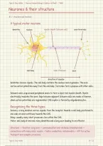
A typical motor neurone
dendrites nucleus myelin sheath (Schwann cell) axon terminals
cell body node of Ranvier axon
direction of impulse
Dendrites receive signals. The cell body contains the nucleus and organelles. The axon
carries action potentials away from the cell body. Terminals form synapses with other cells.
Schwann cells wrap around peripheral axons to form a lipid-rich myelin sheath. Myelin
electrically insulates the axon. Gaps between adjacent Schwann cells are nodes of Ranvier,
where action potentials are regenerated. CNS myelin is formed by oligodendrocytes.
Recognising the three types
Sensory: a long dendron carries signals from the receptor towards a cell body positioned to
one side; an axon continues towards the CNS.
Relay: usually many short processes; lies within the CNS.
Motor: cell body at one end, many dendrites and a long axon leading to an effector.
Structure -> function: long axon = communication over distance; branched ends =
connections with many cells; myelin = faster conduction; mitochondria = ATP for active
transport and synaptic processes.
Topic 8: Grey Matter
,Topic 8: Grey Matter / Pearson Edexcel Biology A (Salters-Nuffield) / 9BN0
Sensory & relay neurones
8.1 / recognising the three types
Sensory neurone
axon terminals
cell body: to one side
direction of impulse
receptor endings dendron: long, towards cell body axon: to the CNS
Relay neurone
cell body
axon terminals
many short dendrites
short axon
the whole neurone lies inside the CNS
Motor neurone
long, myelinated axon
axon terminals at the effector
cell body at one end, with short dendrites
Comparing the three
Sensory Relay Motor
Cell body to one side of the between dendrites and at one end, in the CNS
fibre, outside the CNS axon, in the CNS
Fibres long dendron, shorter short dendrites, short short dendrites, long
axon axon axon
Impulse receptor -> CNS sensory -> motor CNS -> effector
neurone
Topic 8: Grey Matter
, Topic 8: Grey Matter / Pearson Edexcel Biology A (Salters-Nuffield) / 9BN0
Responding to stimuli
8.2 / receptors, effectors and the pupil reflex
A stimulus is a change in the internal or external environment. A receptor detects it; a
coordinator processes the information; an effector produces the response. Muscles
contract and glands secrete.
A spinal withdrawal reflex
Pain stimulus -> receptor in skin -> sensory neurone -> relay neurone in spinal cord ->
motor neurone -> skeletal muscle contracts -> limb withdraws. A reflex is rapid, automatic
and stereotyped. Conscious awareness can occur after the response begins.
cell body in dorsal root ganglion
stimulus
relay neurone
sensory neurone grey matter
receptor (in skin) spinal cord
effector
(muscle)
motor neurone
A spinal reflex is coordinated in the spinal cord. Information can also travel to the brain,
producing conscious awareness of the stimulus. Voluntary responses involve conscious
processing; a protective reflex does not need a conscious decision before the muscles
respond.
Not every reflex has a relay neurone. A simple stretch reflex can connect a sensory
neurone directly to a motor neurone. Synapses introduce a short delay.
Antagonistic muscles in the iris
Bright light: circular muscles contract, radial muscles relax -> pupil constricts -> less
light reaches the retina.
Dim light: radial muscles contract, circular muscles relax -> pupil dilates -> more light
enters. The pupil is an opening; the iris muscles change its diameter.
Pathway: photoreceptors in the retina -> optic nerve -> control centre in the midbrain ->
parasympathetic (oculomotor) nerve -> circular muscles contract. An involuntary reflex.
Topic 8: Grey Matter
Neurones & their structure
8.1 / structure and function
A typical motor neurone
dendrites nucleus myelin sheath (Schwann cell) axon terminals
cell body node of Ranvier axon
direction of impulse
Dendrites receive signals. The cell body contains the nucleus and organelles. The axon
carries action potentials away from the cell body. Terminals form synapses with other cells.
Schwann cells wrap around peripheral axons to form a lipid-rich myelin sheath. Myelin
electrically insulates the axon. Gaps between adjacent Schwann cells are nodes of Ranvier,
where action potentials are regenerated. CNS myelin is formed by oligodendrocytes.
Recognising the three types
Sensory: a long dendron carries signals from the receptor towards a cell body positioned to
one side; an axon continues towards the CNS.
Relay: usually many short processes; lies within the CNS.
Motor: cell body at one end, many dendrites and a long axon leading to an effector.
Structure -> function: long axon = communication over distance; branched ends =
connections with many cells; myelin = faster conduction; mitochondria = ATP for active
transport and synaptic processes.
Topic 8: Grey Matter
,Topic 8: Grey Matter / Pearson Edexcel Biology A (Salters-Nuffield) / 9BN0
Sensory & relay neurones
8.1 / recognising the three types
Sensory neurone
axon terminals
cell body: to one side
direction of impulse
receptor endings dendron: long, towards cell body axon: to the CNS
Relay neurone
cell body
axon terminals
many short dendrites
short axon
the whole neurone lies inside the CNS
Motor neurone
long, myelinated axon
axon terminals at the effector
cell body at one end, with short dendrites
Comparing the three
Sensory Relay Motor
Cell body to one side of the between dendrites and at one end, in the CNS
fibre, outside the CNS axon, in the CNS
Fibres long dendron, shorter short dendrites, short short dendrites, long
axon axon axon
Impulse receptor -> CNS sensory -> motor CNS -> effector
neurone
Topic 8: Grey Matter
, Topic 8: Grey Matter / Pearson Edexcel Biology A (Salters-Nuffield) / 9BN0
Responding to stimuli
8.2 / receptors, effectors and the pupil reflex
A stimulus is a change in the internal or external environment. A receptor detects it; a
coordinator processes the information; an effector produces the response. Muscles
contract and glands secrete.
A spinal withdrawal reflex
Pain stimulus -> receptor in skin -> sensory neurone -> relay neurone in spinal cord ->
motor neurone -> skeletal muscle contracts -> limb withdraws. A reflex is rapid, automatic
and stereotyped. Conscious awareness can occur after the response begins.
cell body in dorsal root ganglion
stimulus
relay neurone
sensory neurone grey matter
receptor (in skin) spinal cord
effector
(muscle)
motor neurone
A spinal reflex is coordinated in the spinal cord. Information can also travel to the brain,
producing conscious awareness of the stimulus. Voluntary responses involve conscious
processing; a protective reflex does not need a conscious decision before the muscles
respond.
Not every reflex has a relay neurone. A simple stretch reflex can connect a sensory
neurone directly to a motor neurone. Synapses introduce a short delay.
Antagonistic muscles in the iris
Bright light: circular muscles contract, radial muscles relax -> pupil constricts -> less
light reaches the retina.
Dim light: radial muscles contract, circular muscles relax -> pupil dilates -> more light
enters. The pupil is an opening; the iris muscles change its diameter.
Pathway: photoreceptors in the retina -> optic nerve -> control centre in the midbrain ->
parasympathetic (oculomotor) nerve -> circular muscles contract. An involuntary reflex.
Topic 8: Grey Matter


