Cytology: is the study of the cell.
Cytopathology: the science dealing with the structure of abnormal or diseased cell Cytology specimens
- A cell is the basic structural, functional and biological unit of a living organism 1. Peritoneal, pericardial and pleural fluids
- Tissues consist entirely of cells and extracellular matrix elaborated by cells. 2. CSF
- Most mammalian cells are microscopic 3. Nipple discharge
4. Bronchial brushings / washings
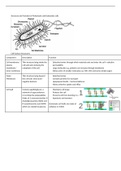
Types of microscopes: 5. Sputum
- Light microscopy 6. Gastric washings
- Fluorescence microscopy 7. Urine sediment
- Confocal microscopy 8. Prostatic secretions
- Electron microscopy 9. Cervicovaginal (paps) smear
Cytology Sampling Techniques 1. Pap smear - Papanicolaou Staining Method (pap staining):
- Exfoliative cytology: cells shed/scraped/brushed off an epithelial a. Developed by Dr. George N. Papanicolaou
surface b. Polychrome staining reaction (Haematoxylin, Orange G ,
- Fluid cytology: cells withdrawn with the fluid in which they are Eosin, Eosin Y, Light Green SF yellowish, and Bismarck
suspended brown Y)
- Washings: cells flushed out of an organ using an irrigating fluid c. Display the many variations of cellular morphology
- Fine –needle aspiration cytology: cells sucked out of a solid tissue d. Mainly for cervical cancer
using a thin needle attached to a syringe. 2. Hematoxylin/Eosin staining
a. Hydration: (nuclear dye, Haematoxylin, blue).
Examining Cytology Samples: b. Dehydration: (counterstains, cytoplasm (Eosin, pink).
1. Pap Staining c. Clearing
2. Hematoxylin/Eosin (H & E) d. Mounting
3. Immunocytochemistry 3. Immunocytochemistry
a. Detection of surface antigens (markers) on isolated cells
Cytology – specimen processing: Common fixatives: b. The detection is based on specific antigen antibody binding
- 95% Ethyl alcohol (immune reactions).
- 100% Ethanol c. A specific antibody identifies an epitope with 8-15 length
- 100% methanol amino acid sequence in
- 80% isopropanol d. A protein (epitopes).
Immunocytochemistry:
- Tumour diagnostic/classification
- Prognostic/Predictor Markers
- Target Therapy
Homogenisation and fractionation
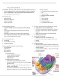
- Cell is the structure and functional unit of life
- Cell contain organelles which perform a variety of biological
specific functions
- Electron micrograph explains only the structure but not the
function of cell organelles
- To obtain precise information about the cells organelles, it is
necessary to isolate them free from contamination organelles
Cytopathology: the science dealing with the structure of abnormal or diseased cell Cytology specimens
- A cell is the basic structural, functional and biological unit of a living organism 1. Peritoneal, pericardial and pleural fluids
- Tissues consist entirely of cells and extracellular matrix elaborated by cells. 2. CSF
- Most mammalian cells are microscopic 3. Nipple discharge
4. Bronchial brushings / washings
Types of microscopes: 5. Sputum
- Light microscopy 6. Gastric washings
- Fluorescence microscopy 7. Urine sediment
- Confocal microscopy 8. Prostatic secretions
- Electron microscopy 9. Cervicovaginal (paps) smear
Cytology Sampling Techniques 1. Pap smear - Papanicolaou Staining Method (pap staining):
- Exfoliative cytology: cells shed/scraped/brushed off an epithelial a. Developed by Dr. George N. Papanicolaou
surface b. Polychrome staining reaction (Haematoxylin, Orange G ,
- Fluid cytology: cells withdrawn with the fluid in which they are Eosin, Eosin Y, Light Green SF yellowish, and Bismarck
suspended brown Y)
- Washings: cells flushed out of an organ using an irrigating fluid c. Display the many variations of cellular morphology
- Fine –needle aspiration cytology: cells sucked out of a solid tissue d. Mainly for cervical cancer
using a thin needle attached to a syringe. 2. Hematoxylin/Eosin staining
a. Hydration: (nuclear dye, Haematoxylin, blue).
Examining Cytology Samples: b. Dehydration: (counterstains, cytoplasm (Eosin, pink).
1. Pap Staining c. Clearing
2. Hematoxylin/Eosin (H & E) d. Mounting
3. Immunocytochemistry 3. Immunocytochemistry
a. Detection of surface antigens (markers) on isolated cells
Cytology – specimen processing: Common fixatives: b. The detection is based on specific antigen antibody binding
- 95% Ethyl alcohol (immune reactions).
- 100% Ethanol c. A specific antibody identifies an epitope with 8-15 length
- 100% methanol amino acid sequence in
- 80% isopropanol d. A protein (epitopes).
Immunocytochemistry:
- Tumour diagnostic/classification
- Prognostic/Predictor Markers
- Target Therapy
Homogenisation and fractionation
- Cell is the structure and functional unit of life
- Cell contain organelles which perform a variety of biological
specific functions
- Electron micrograph explains only the structure but not the
function of cell organelles
- To obtain precise information about the cells organelles, it is
necessary to isolate them free from contamination organelles