Communicable
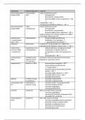
Primary defences
diseases Secondary defences
The skin (main primary defence) Antigens
- Epidermis- outer layer of the skin - Chemical markers on the outer membrane of
1. Keratinocytes produced by mitosis at the base of pathogens intrinsic proteins to the plasma
the epidermis membrane
Opsonins
2. They migrate to the surface of the skin
- Protein molecules that attach to antigens on
3. As they migrate the cells dry out, so the
the surface of a pathogen, it is a type of
cytoplasm is converted into keratin
antibody to enhance ability of phagocytic
4. When the cells do reach the surface, they are
cells to bind and engulf pathogens
dead, dead cells act as a barrier to infection.
Phagocytes
Neutrophils Macrophages
Mucous membranes Engulfs foreign matter Travel in the blood as
and traps it inside a monocytes to then
- Exchange surfaces are thinner for a short diffusion
large vacuole engulf foreign matter to
distance but less protected from pathogens. trap it in a large vacuole
- Epithelial layer contains: mucus-secreting cells and Multilobed Kidney shaped
goblet cells nucleus nucleus
- Mucus-secreting cells= lines the passages to trap Many lysosomes Larger nucleus
pathogens Released in large Travel in the
- Goblet cells= hair like and can move, to waft layers of numbers blood as
mucus containing pathogens to the top of the monocytes
trachea to be swallowed and digested 1. Bind to opsonin 1. Bind to opsonin
attached to antigen attached to antigen
2. Engulfs pathogen 2. Engulfs pathogen by
by endocytosis to endocytosis to form
Inflammation form phagosome phagosome
1. Mast cells detect the presence of microorganisms 3. lysosome fuses with 3. lysosome fuses with
2. They then release histamine which causes phagosome to form phagosome to form
phagolysosome phagolysosome
vasodilation
4. releases lytic 4. releases lytic
3. White blood cells leave blood and become part of enzymes to be enzymes, pathogen
the tissue fluid digested and only is not fully digested
4. Excess tissue fluid is then drained into the the harmless so moves antigen on
lymphatic system products are the surface to
5. Pathogens encounter lymphocytes and initiate the absorbed into the protein complex on
specific immune response. cell cell surface to be
presented
Vasodilation = making capillary walls more permeable
to white blood cells and proteins
Blood clotting
1. Platelets release clotting factors which activate
the enzyme cascade
2. A scab forms and the skin repairs underneath
3. Fibrous collagen is deposited
4. Stem cells undergo mitosis and migrate towards
the skins surface
5. New cells supply oxygen to skin tissues, they then
Communicable
can contact to draw edges of the cut together
6. New skin is completed and the scab is released
diseases
The specific immune response
B lymphocytes (made and
5. Macrophages
matured in the Bone marrow)
present antigens
on their surface T lymphocytes (made in
bone marrow and matured in
the Thymus)