CLINICAL IMMUNOLOGY
TOPIC 2: IBD & CELIAC DISEASE
LECTURE 1: MOLECULAR BASIS OF CELIAC DISEASE Wednesday, 13/11/2019
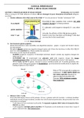
What is wrong in CD? With influence of wheat, villi are damaged → poor absorption of nutrients (diagram
see slide)
a. Genetic influence: HLA-DQ2 and/or HLA-DQ8 → *is it also present in “healthy” individuals? YUP.
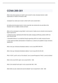
HLA-DQ2/8 likes peptides that contain AA with
negative charge in certain positions
E = glutamic acid (negative-charged); X = any other
AA
Actually, the affinity of HLA-DQ/gluten peptide
bonds is very low, but there are hecking T cells that
are reactive to this weak bond
b. Environment: gluten peptides
Gluten characteristics: water-insoluble, non-digestible (amylase – pepsin – trypsin can’t break it down)
for some people
But gluten doesn’t contain any negative-charged amino acid → how the heck does it affect HLA-DQ2/8
then? It is hella modified hey. What modifies the molecule?
First, these are the important peptides:
- GLIADIN (fragment of pepsin) → HLA-DQ8 restricted; doesn’t contain any negative-charged AA
either (no E’s)
- GLUTAMINE (Q) → transformed into glutamic acid with negative charge (by tissue
transglutaminase/TTG); exerts the noxious effect; Q→E modification exerts T cell response
TTG is highly specific to gluten, released in stress condition/acidic pH → modify glutamine into
glutamic acid (creating new epitope by deamidation – posttranslational modification) & amplify the
binding of gluten to HLA-DQ2/8
Why is TTG very specific to gluten in
intestines? → QXP/QXPY/QXPF are
the tissue-specific sequence of
gluten-induced intestinal villi
damage, 30% of gluten contain
that sequence
c. Effector: CD4+ T cells
From intestinal biopsy tissues of CD patients → Case = patients with CD; Controls = relatively healthier
individuals, mostly not age-matched; consider the risk of intestinal perforation, thus procedure has to be
undertaken accordingly
- T cell clones 2/5 are restricted via HLA-DQ → responds to gluten ONLY when it’s bound to HLA-
DQ2/8 (see slide)
- T cell recognizes multiple gluten peptides → CD patients can be intolerant to more than 1 type of
gluten; Characteristic: PRO-inflammatory
, CLINICAL IMMUNOLOGY
TOPIC 2: IBD & CELIAC DISEASE
- Gluten-reactive T cells are primed after the child is weaned/more solid food is introduced → various
T cells reacting to various gluten epitopes are generated early in life, some can break the individual’s
tolerance to gluten
d. Impact: tissue damage in intestines
*How does some people realize their gluten
intolerance later in life if the priming occurs very
early in life? → Not everyone has similar symptoms,
tolerance is varied among individuals - mild
symptoms can be ignored/misdiagnosed
Conclusion: there is a PERFECT BUT FATAL
MATCH between HLA-DQ molecule and TTG-
modified gluten → causing tissue damage
Paradox: 95% of patients is HLA-DQ2 (+) but 95% of HLA-DQ2 (+) individuals DO NOT DEVELOP CD; WHY?
a. High affinity T cell responses to immunodominant gluten peptides (gluten-reactive T cells) are only
found in CD patients
b. T cell repertoire among CD patients (as well as non-CD individuals) are unique to the individual → no
one is 100% the same, even among CD patients themselves, however, there are some some similarity
among the individuals with CD
- TRAV26-BV9 bias → …?
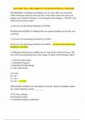
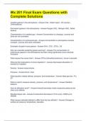
- CDR3 alfa/CDR3 beta regions of the T cell receptors are the same in 2 different CD patients; Arginine
in the middle of CDR3 alfa sequence (inserted later, non-germline) → arginine is in contact with
gluten particle, induce the T cell recognition of gluten peptide as foreign
If arginine is replaced/removed → no recognition by gluten-reactive T cell
Blue = gluten, grey = HLA
- Some people’s T cell are not primed to react against gluten in thymus → may explain why some
people don’t develop intolerance to gluten at all
c. Deamidated gluten peptide induce B cell response → create auto-antibodies
Conclusion: biased TCR repertoire is
structurally conserved → expansion of T
cells is necessary for CD development
*Does it impact Dx & Tx? PROBABLY
, CLINICAL IMMUNOLOGY
TOPIC 2: IBD & CELIAC DISEASE
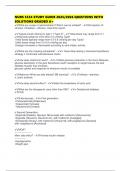
Mass cytometry (next gen flowcytometry) can work with multiple markers (>12 markers) simultaneously w/o
spectral overlap
a. Benefit: more extensive, dimensionality reduction techniques possible
Visible markers: CD3, CD7
Other benefits: localize T cells, differentiate various cells in biopsy sample
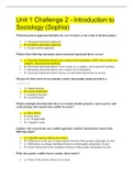
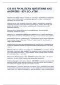
b. t-SNE → …?
Grouping the cells based on their similarities; green = Treg, others = conventional T cells
Through t-SNE we can see that gluten-specific CD4+ T cells are present since early life & 28 distinct
subsets (for …?) in gluten-specific CD4+ T cells especially in gastrointestinal organs (when analysis is
combined with heat map)
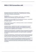
t-SNE can also distinguish the source of samples → healthy control/patients, not just the location from
which the sample is taken
c. unknown cells between NK cells & ILCs → plastic, it differentiates into NK cells/ILC depending on the
administered interleukins
TOPIC 2: IBD & CELIAC DISEASE
LECTURE 1: MOLECULAR BASIS OF CELIAC DISEASE Wednesday, 13/11/2019
What is wrong in CD? With influence of wheat, villi are damaged → poor absorption of nutrients (diagram
see slide)
a. Genetic influence: HLA-DQ2 and/or HLA-DQ8 → *is it also present in “healthy” individuals? YUP.
HLA-DQ2/8 likes peptides that contain AA with
negative charge in certain positions
E = glutamic acid (negative-charged); X = any other
AA
Actually, the affinity of HLA-DQ/gluten peptide
bonds is very low, but there are hecking T cells that
are reactive to this weak bond
b. Environment: gluten peptides
Gluten characteristics: water-insoluble, non-digestible (amylase – pepsin – trypsin can’t break it down)
for some people
But gluten doesn’t contain any negative-charged amino acid → how the heck does it affect HLA-DQ2/8
then? It is hella modified hey. What modifies the molecule?
First, these are the important peptides:
- GLIADIN (fragment of pepsin) → HLA-DQ8 restricted; doesn’t contain any negative-charged AA
either (no E’s)
- GLUTAMINE (Q) → transformed into glutamic acid with negative charge (by tissue
transglutaminase/TTG); exerts the noxious effect; Q→E modification exerts T cell response
TTG is highly specific to gluten, released in stress condition/acidic pH → modify glutamine into
glutamic acid (creating new epitope by deamidation – posttranslational modification) & amplify the
binding of gluten to HLA-DQ2/8
Why is TTG very specific to gluten in
intestines? → QXP/QXPY/QXPF are
the tissue-specific sequence of
gluten-induced intestinal villi
damage, 30% of gluten contain
that sequence
c. Effector: CD4+ T cells
From intestinal biopsy tissues of CD patients → Case = patients with CD; Controls = relatively healthier
individuals, mostly not age-matched; consider the risk of intestinal perforation, thus procedure has to be
undertaken accordingly
- T cell clones 2/5 are restricted via HLA-DQ → responds to gluten ONLY when it’s bound to HLA-
DQ2/8 (see slide)
- T cell recognizes multiple gluten peptides → CD patients can be intolerant to more than 1 type of
gluten; Characteristic: PRO-inflammatory
, CLINICAL IMMUNOLOGY
TOPIC 2: IBD & CELIAC DISEASE
- Gluten-reactive T cells are primed after the child is weaned/more solid food is introduced → various
T cells reacting to various gluten epitopes are generated early in life, some can break the individual’s
tolerance to gluten
d. Impact: tissue damage in intestines
*How does some people realize their gluten
intolerance later in life if the priming occurs very
early in life? → Not everyone has similar symptoms,
tolerance is varied among individuals - mild
symptoms can be ignored/misdiagnosed
Conclusion: there is a PERFECT BUT FATAL
MATCH between HLA-DQ molecule and TTG-
modified gluten → causing tissue damage
Paradox: 95% of patients is HLA-DQ2 (+) but 95% of HLA-DQ2 (+) individuals DO NOT DEVELOP CD; WHY?
a. High affinity T cell responses to immunodominant gluten peptides (gluten-reactive T cells) are only
found in CD patients
b. T cell repertoire among CD patients (as well as non-CD individuals) are unique to the individual → no
one is 100% the same, even among CD patients themselves, however, there are some some similarity
among the individuals with CD
- TRAV26-BV9 bias → …?
- CDR3 alfa/CDR3 beta regions of the T cell receptors are the same in 2 different CD patients; Arginine
in the middle of CDR3 alfa sequence (inserted later, non-germline) → arginine is in contact with
gluten particle, induce the T cell recognition of gluten peptide as foreign
If arginine is replaced/removed → no recognition by gluten-reactive T cell
Blue = gluten, grey = HLA
- Some people’s T cell are not primed to react against gluten in thymus → may explain why some
people don’t develop intolerance to gluten at all
c. Deamidated gluten peptide induce B cell response → create auto-antibodies
Conclusion: biased TCR repertoire is
structurally conserved → expansion of T
cells is necessary for CD development
*Does it impact Dx & Tx? PROBABLY
, CLINICAL IMMUNOLOGY
TOPIC 2: IBD & CELIAC DISEASE
Mass cytometry (next gen flowcytometry) can work with multiple markers (>12 markers) simultaneously w/o
spectral overlap
a. Benefit: more extensive, dimensionality reduction techniques possible
Visible markers: CD3, CD7
Other benefits: localize T cells, differentiate various cells in biopsy sample
b. t-SNE → …?
Grouping the cells based on their similarities; green = Treg, others = conventional T cells
Through t-SNE we can see that gluten-specific CD4+ T cells are present since early life & 28 distinct
subsets (for …?) in gluten-specific CD4+ T cells especially in gastrointestinal organs (when analysis is
combined with heat map)
t-SNE can also distinguish the source of samples → healthy control/patients, not just the location from
which the sample is taken
c. unknown cells between NK cells & ILCs → plastic, it differentiates into NK cells/ILC depending on the
administered interleukins