2, 3 (3.1, 3.5-3.10)
17 jan 2024 Lecture 6 (Chapter 2)
2.1 The Cytoplasmic Membrane
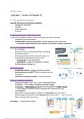
Layered structures surrounding cytoplasm:
- cytoplasmic membrane
- cell wall
- outer membrane
- S-layers
Cytoplasmic Membrane / Plasma Membrane
= surrounds cytoplasm (mixture of macromolecules and small molecules)
- separates it from environment
- main function: selective permeability (nutrient transport in and waste out)
→ membrane proteins facilitate these reactions and function in energy metabolism
Bac + Euk Cytoplasmic Membranes
- 8-10 nm wide
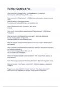
- general structure is phospholipid bilayer containing
embedded proteins
- Containing both hydrophilic (water-attracting) and
hydrophobic (water-repellent) components
→ hydrophobic = fatty acids (tails)
→ hydrophilic = glycerol + phosphate + another
functional group (sugars e.g.)
Membrane proteins
- embedded proteins = integral membrane proteins
- transmembrane proteins = extend completely across
membrane
- peripheral membrane proteins = loosely attached
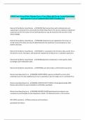
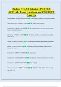
Archaea Cytoplasmic Membrane
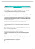
- structure is similar to bac + euk, but it is chemically different
- In bac/Euk → FA are bound to glycerol via ester linkages
In Archaea → Isoprenoids (ipv FA) are bound to glycerol via ether linkages
→ many different isoprenoid chains including some ring structures (e.g.
crenarchaeol)
- Major lipids are phosphoglycerol diethers with phytanyl (C20) side chains +
diphosphoglycerol tetraethers with biphytanyl (C40) side chains → which can form
lipid monolayers (fig c) ipv bilayer
monolayer → isoprenoids are linked
,Cytoplasmic Membrane Function:
1. Permeability barrier
- polar and charged molecules must be transported
- transport proteins accumulate solutes against the concentration gradient
- prevent leakage
2. Protein anchor
- holds proteins in place
3. Energy conservation and consumption
- generation of proton motive force (potential energy present)
Membrane transport:
solute = particle
2.2 Transporting nutrients into the cell
Active transport = how cells accumulate solutes against concentration gradient
Transporters
= energy-driven (proton motive force, ATP, or another
energy rich compound)
3 mechanisms
- simple transport = transmembrane transport
protein
→ driven by pmf , noATP
→ Symport (one direction) or Antiport (2 solutes
transported in opposite direction)
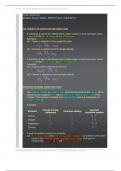
- group translocation = series of proteins
→ substance transported is chemically modified
→ energy rich organic compound (not pmf) drives transport
- ABC system = 3 components (binding protein, transmembrane transporter,
ATP-hydrolyzing protein)
,Group translocation
● Phosphotransferase system in E coli
- best studied translocation system
- glucose, fructose, mannose
- 5 proteins required
- energy from phosphoenolpyruvate (from glycolysis)
ABC-transporter systems
- ABC = ATP-binding cassette
- 200+ different systems for organic and inorganic compounds
- substrate -binding proteins outside of the cell have high substrate affinity
- ATP drives uptake
2.3 The Cell Wall
● Needs to withstand osmotic/turgor pressure to prevent cell lysis
● Maintains cell shape and rigidity
● Most Bacteria separated into 2 groups based on gram-stain (organization and cell
wall structures
Gram-stain
● gram-positive and gram-negatives have different cell wall structures
- gram positive cell envelope
→ cytoplasmic membrane + thick cell wall
- gram negative cell envelope
→ cytoplasmic membrane + thin cell wall + outer membrane + periplasm
● gram stain reaction determined by cell wall thickness
Bacterial cell wall
- Peptidoglycan = rigid polysaccharide layer that provides strength (component of cell
wall)
→ not found in archaea and eukaryotic cells
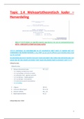
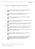
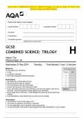
- Glycan tetrapeptide (peptidoglycan) contains:
, - Sugar backbone of peptidoglycan is composed of alternating
repeats of two modified glucose residues called
N-acetylglucosamine and N-acetylmuramic acid joined by a b-1,4
linkage
- Short peptide attached to N-acetylmuramic acid
→ amino acids vary between species
→ Amino acids are: L-alanine, D-alanine, D-glutamic acid and
L-lysine of diaminopimelic acid (DAP)
● Peptidoglycan strands run parallel around cell circumference (pic)
● strands are cross-linked by covalent peptide bonds (a)
- becomes one big molecule
● gram-negative crosslinks between DAP amino and D-alanine
carboxyl on adjacent glycan strand
- primarily single layer
● peptidoglycan mesh formed is flexible and porous → but strong
enough to resist turgor pressure and prevent rupture
- additional strength in gram negative → provided by outer
membrane
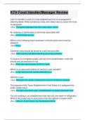
Gram-positive cell envelope
- Thick peptidoglycan cell wall (20-35 nm)
- Up to 90% peptidoglycan (15> layers)
- stabilized by horizontal and vertical peptide cross-links often
containing peptide interbridges
- Commonly have teichoic acids (acidic molecules) embedded in cell wall and
covalently linked to peptidoglycan
→ lipoteichoic acids: teichoic acids covalently bound to membrane lipids
- Peptidoglycan can be destroyed by lysozyme ⇒ cleaves
glycosidic bond between sugars
→ major defense against bacterial infection
- Penicillin blocks formation of peptide cross-links
- ⇒ picture ⇒
Archaea walls
- Cytoplasmic membrane different from Bac
- Lack peptidoglycan
- typically lack outer membrane → gram staining does not work
- most lack polysaccharide wall ⇒ instead they have S-layer
(protein shell) → prevents osmotic lysis
● Cell walls have unique chemical structures (not found in Bac)
17 jan 2024 Lecture 6 (Chapter 2)
2.1 The Cytoplasmic Membrane
Layered structures surrounding cytoplasm:
- cytoplasmic membrane
- cell wall
- outer membrane
- S-layers
Cytoplasmic Membrane / Plasma Membrane
= surrounds cytoplasm (mixture of macromolecules and small molecules)
- separates it from environment
- main function: selective permeability (nutrient transport in and waste out)
→ membrane proteins facilitate these reactions and function in energy metabolism
Bac + Euk Cytoplasmic Membranes
- 8-10 nm wide
- general structure is phospholipid bilayer containing
embedded proteins
- Containing both hydrophilic (water-attracting) and
hydrophobic (water-repellent) components
→ hydrophobic = fatty acids (tails)
→ hydrophilic = glycerol + phosphate + another
functional group (sugars e.g.)
Membrane proteins
- embedded proteins = integral membrane proteins
- transmembrane proteins = extend completely across
membrane
- peripheral membrane proteins = loosely attached
Archaea Cytoplasmic Membrane
- structure is similar to bac + euk, but it is chemically different
- In bac/Euk → FA are bound to glycerol via ester linkages
In Archaea → Isoprenoids (ipv FA) are bound to glycerol via ether linkages
→ many different isoprenoid chains including some ring structures (e.g.
crenarchaeol)
- Major lipids are phosphoglycerol diethers with phytanyl (C20) side chains +
diphosphoglycerol tetraethers with biphytanyl (C40) side chains → which can form
lipid monolayers (fig c) ipv bilayer
monolayer → isoprenoids are linked
,Cytoplasmic Membrane Function:
1. Permeability barrier
- polar and charged molecules must be transported
- transport proteins accumulate solutes against the concentration gradient
- prevent leakage
2. Protein anchor
- holds proteins in place
3. Energy conservation and consumption
- generation of proton motive force (potential energy present)
Membrane transport:
solute = particle
2.2 Transporting nutrients into the cell
Active transport = how cells accumulate solutes against concentration gradient
Transporters
= energy-driven (proton motive force, ATP, or another
energy rich compound)
3 mechanisms
- simple transport = transmembrane transport
protein
→ driven by pmf , noATP
→ Symport (one direction) or Antiport (2 solutes
transported in opposite direction)
- group translocation = series of proteins
→ substance transported is chemically modified
→ energy rich organic compound (not pmf) drives transport
- ABC system = 3 components (binding protein, transmembrane transporter,
ATP-hydrolyzing protein)
,Group translocation
● Phosphotransferase system in E coli
- best studied translocation system
- glucose, fructose, mannose
- 5 proteins required
- energy from phosphoenolpyruvate (from glycolysis)
ABC-transporter systems
- ABC = ATP-binding cassette
- 200+ different systems for organic and inorganic compounds
- substrate -binding proteins outside of the cell have high substrate affinity
- ATP drives uptake
2.3 The Cell Wall
● Needs to withstand osmotic/turgor pressure to prevent cell lysis
● Maintains cell shape and rigidity
● Most Bacteria separated into 2 groups based on gram-stain (organization and cell
wall structures
Gram-stain
● gram-positive and gram-negatives have different cell wall structures
- gram positive cell envelope
→ cytoplasmic membrane + thick cell wall
- gram negative cell envelope
→ cytoplasmic membrane + thin cell wall + outer membrane + periplasm
● gram stain reaction determined by cell wall thickness
Bacterial cell wall
- Peptidoglycan = rigid polysaccharide layer that provides strength (component of cell
wall)
→ not found in archaea and eukaryotic cells
- Glycan tetrapeptide (peptidoglycan) contains:
, - Sugar backbone of peptidoglycan is composed of alternating
repeats of two modified glucose residues called
N-acetylglucosamine and N-acetylmuramic acid joined by a b-1,4
linkage
- Short peptide attached to N-acetylmuramic acid
→ amino acids vary between species
→ Amino acids are: L-alanine, D-alanine, D-glutamic acid and
L-lysine of diaminopimelic acid (DAP)
● Peptidoglycan strands run parallel around cell circumference (pic)
● strands are cross-linked by covalent peptide bonds (a)
- becomes one big molecule
● gram-negative crosslinks between DAP amino and D-alanine
carboxyl on adjacent glycan strand
- primarily single layer
● peptidoglycan mesh formed is flexible and porous → but strong
enough to resist turgor pressure and prevent rupture
- additional strength in gram negative → provided by outer
membrane
Gram-positive cell envelope
- Thick peptidoglycan cell wall (20-35 nm)
- Up to 90% peptidoglycan (15> layers)
- stabilized by horizontal and vertical peptide cross-links often
containing peptide interbridges
- Commonly have teichoic acids (acidic molecules) embedded in cell wall and
covalently linked to peptidoglycan
→ lipoteichoic acids: teichoic acids covalently bound to membrane lipids
- Peptidoglycan can be destroyed by lysozyme ⇒ cleaves
glycosidic bond between sugars
→ major defense against bacterial infection
- Penicillin blocks formation of peptide cross-links
- ⇒ picture ⇒
Archaea walls
- Cytoplasmic membrane different from Bac
- Lack peptidoglycan
- typically lack outer membrane → gram staining does not work
- most lack polysaccharide wall ⇒ instead they have S-layer
(protein shell) → prevents osmotic lysis
● Cell walls have unique chemical structures (not found in Bac)