Course 2.4 Brain and Cognition: Perception
Problem 1: A Keen Eye
Literature: Wolfe, Coren
A little about light:
- Travels in waves, “photons” when absorbed
- Visible wavelength: 400nm (violet) – 6500nm (red)
- Visual system interprets color from wavelength
- Absorption = taking up light
- Reflection = bouncing off (e.g. from light colored surface)
- Transmission = waves go through surface
- Refraction = waves are bent
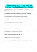
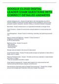
Anatomy of the eye:
Name Short description/function
A Vitreous body Space between retina and lens. 80% of internal eye volume. Gel-like viscous (like egg white),
(humor) generally transparent. “Floaters” = bio debris that drifts around.
B Optic nerve
C Fovea High acuity. Use to identify objects, read, look at details.
D Retina Where light waves are brought into focus. Detects light and tells brain. Beginning of
“seeing”.
E Choroid
F Sclera Strong elastic membrane. Seen as “white” of the eye
G Cornea Highly curved transparent “window to the world”. No blood (vessels). Has a lot of
transparent nerve endings (→ force tears when cornea is scratched). Can regenerate/heal
quickly (24hrs). High refractive index that can change due to lens (→bends light)
H Anterior Space right behind cornea filled with fluid. No blood. Brings nutrients and oxygen to cornea
chamber and lens. Removes waste.
I Pupil Light needs to pass through pupil to get to the lens. A hole in the iris. Controls amount of
light that reaches retina (= pupillary light reflex).
Automatic expansion/contraction depending on light (Whytt´s reflex)
J Iris Gives eye color. Muscular diaphragm around pupil which regulates contraction/expansion
of it.
K (Crystalline) Transparent (= no blood supply). // slightly yellow; tint increases with age. Controlled by
Lens ciliary muscle. Can change shape and focus on things (=accommodation). Attached to ciliary
muscle; relaxed → flat lens → distant objects; tense → lens bulges → close objects. Fat lens
= more power
L Blind spot/ Where axons of ganglion cells leave eye through optic nerve. Veins and arteries. No
optic disc photoreceptors, therefore blind.
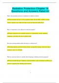
The retina:
- Human retina = duplex retina
- Light energy transduced into neural energy
- Fundus = back surface of the eye
- Optic disc/ blind spot usually not notice because the visual system fills it in
- Neurons connect to front most layer of retina → optic nerve → brain
- Pigment epithelium = backs retina. Dark, pigmented layer to stop light from reflecting. Reflected light causes
blurring/fogging.
- Nocturnal animals need more light detection therefore retina is backed with reflecting tapetum (light passes
through retina twice)
o Better light sensitivity; but image is blurry
Problem 1: A Keen Eye
Literature: Wolfe, Coren
A little about light:
- Travels in waves, “photons” when absorbed
- Visible wavelength: 400nm (violet) – 6500nm (red)
- Visual system interprets color from wavelength
- Absorption = taking up light
- Reflection = bouncing off (e.g. from light colored surface)
- Transmission = waves go through surface
- Refraction = waves are bent
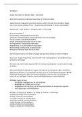
Anatomy of the eye:
Name Short description/function
A Vitreous body Space between retina and lens. 80% of internal eye volume. Gel-like viscous (like egg white),
(humor) generally transparent. “Floaters” = bio debris that drifts around.
B Optic nerve
C Fovea High acuity. Use to identify objects, read, look at details.
D Retina Where light waves are brought into focus. Detects light and tells brain. Beginning of
“seeing”.
E Choroid
F Sclera Strong elastic membrane. Seen as “white” of the eye
G Cornea Highly curved transparent “window to the world”. No blood (vessels). Has a lot of
transparent nerve endings (→ force tears when cornea is scratched). Can regenerate/heal
quickly (24hrs). High refractive index that can change due to lens (→bends light)
H Anterior Space right behind cornea filled with fluid. No blood. Brings nutrients and oxygen to cornea
chamber and lens. Removes waste.
I Pupil Light needs to pass through pupil to get to the lens. A hole in the iris. Controls amount of
light that reaches retina (= pupillary light reflex).
Automatic expansion/contraction depending on light (Whytt´s reflex)
J Iris Gives eye color. Muscular diaphragm around pupil which regulates contraction/expansion
of it.
K (Crystalline) Transparent (= no blood supply). // slightly yellow; tint increases with age. Controlled by
Lens ciliary muscle. Can change shape and focus on things (=accommodation). Attached to ciliary
muscle; relaxed → flat lens → distant objects; tense → lens bulges → close objects. Fat lens
= more power
L Blind spot/ Where axons of ganglion cells leave eye through optic nerve. Veins and arteries. No
optic disc photoreceptors, therefore blind.
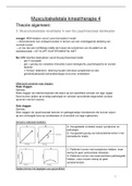
The retina:
- Human retina = duplex retina
- Light energy transduced into neural energy
- Fundus = back surface of the eye
- Optic disc/ blind spot usually not notice because the visual system fills it in
- Neurons connect to front most layer of retina → optic nerve → brain
- Pigment epithelium = backs retina. Dark, pigmented layer to stop light from reflecting. Reflected light causes
blurring/fogging.
- Nocturnal animals need more light detection therefore retina is backed with reflecting tapetum (light passes
through retina twice)
o Better light sensitivity; but image is blurry