Neurocognition
Lecture 1 – The brain and cognition over the life spam – 5/9/22
Brain structure & anatomy
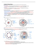
Various types of neural and glia cells classified based on their shape, location and function:
Neurons: different cell types categorized by shape and function
- Sensory (afferent = towards the brain)
- Interneurons (stellate, pyramidal, pukinje)
- Motor (efferent = away from the brain) – sending out motor functions
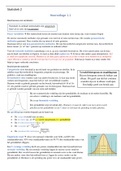
Neurons communicate through action potentials and synaptic transfer
Action potentials – sending these signals
- Thresholded, non-decremental, all-or-nothing response
- What can vary? Rhytm, how many potentials are being fired off
- Triggered by summation of excitatory potentials
- Driven by varying ion permeability of cell membrane
- Propagates along axon (can travel for a meter or more)
- Triggers neurotransmitter release at axon terminal
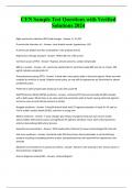
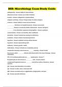
Synapse:
Action potentials leads to neurotransmitter release into synaptic cleft:
- Some neurons can release more than one type of NT, depending on type of stimulation (low vs. high
frequency stimulation): Acetylcholine, dopamine, norepinephrine, serotonin, glutamate, GABA (few)
- Receptor cells in the postsynaptic membrane can adapt to under- or overuse
- The distribution of synapses connecting to a cell influences its excitability (summation, whether an
axtion potential fires or not)
Glia cells
Vary in shape, size and function
- Astrocytes: help with structural support and blood-brain barrier
- Oligodendrocytes: myelin (allows action potential to go faster) for CNS (central nervous system)
neurons
- Microglial cells: fight infections, waste disposal
,- Ependymal cells: ventricular surface epithelium, create CSF (carinal spinal flued)
- Schwann cells: myelin for peripheral neurons (so outside of the brain)
Grey matter consists of neuronal cell bodies and glia cells, situated on the outer layer of the
hemisphere (cortex) and in subcortical nuclei
Cortical cell layers (cortex – outside layers of the hemisphere)
- Different type of neuron (cells) are often organized in layers
- Sensory (input), interneurons (relay) and motor (output) neurons are grouped
- Layers are different in different crotical areas, depending on primary function
White matter consists of neuronal axons and glia cells, organized in bundles
that connects different grey matter areas
White matter tracs – bundles of myelinated axons (myelin looks white)
Connecting neurons throughout the central and peripheral nervous system
- Association fibers connecting areas within a hemisphere (same side)
- Commissural fibers crossing to the other hemisphere,
to the same (homotopic) or a different place (heterotopic)
- Projection fibers connect outwards, to subcortical regions, cerebellum
or the spinal chord
→ different types of connections – sending signals outwards or in brain
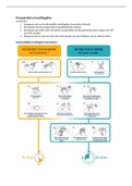
Main components of the brain can be identified and subdivided into specific areas:
Major components of the CNS
- Forebrains: incl hemispheres, corpus callosum and subcortical deep structures (telencephalon)
- Diencephalon: incl thalamic structures
- Midbrain (mesencephalon) - top of brain stem; incl motor and sensory relay nuclei
- Hindbrain (metencephalon); incl pons and cerebellum, medulla oblongata
- Spinal chord
Hindbrain and midbrain:
Hindbrain:
- Medulla oblongata: contributes to vital reflexes, damage is often fatal
- Pons: Crossing over of many fibers in the motor and sensory pathways for contralateral motor
control
- Cerebellum: automated movement, balance, timing & time perfection, sensorimotor coupling,
attention shifting, other cognitive functions
- Origin of cranial nerves V-XII:
Midbrain/ mesecephalon:
- Superior/ inferior colliculi: contribute to sensory processing
- Substantia nigra: contributes to movement initiation
- Origin of cranial nerves II-IV
,Diencephalon:
- Thalamus: important relay station (many subnuclei); any information that goes in or out passes
through the thalamus at some point → delivers signals to almost the whole brain
- Hypothalamus, pituary gland
Some areas are highly specialized, and some areas are just a relay station/ gating functions
Thalamus:
Telencephalon (or cerebrum): subcortical areas: anything underneath the cortex
- Thalamus
- Basal ganglia (see underneath): located deep in the brain
- Limbic structures
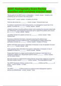
Basal ganglia circuits:
Multiple circuits in the brain go through the basal ganglia:
- Motor circuit: organizing voluntary movement through inhibitory excitatory pathways
- Associative ciruit: contributing to learning, predictive processing, sequencing (sequence of
movements/ learning)
- Reward circuit: producing pleasure responses, motivational functions
→ circuits have functionally relation to each other: reward is important for learning etc
Limbic system (stress response, emotional processing):
- Cingulate (part of cortex, so not entirely subcortical)
- Hippocampus
- Hypothalamus
- Amygdala
→ emotional coloring of what you experience
, Telencephalon: cortical lobes (not the cerebellum):
- Frontal: EF, planning, impulse control, motor function, reward circuit, short-term memory etc
→ mainly about output, behavior, how we manage ourself in the world
- Parietal: sensory integration, association processes, language functions, spatial processing, sense of
touch, some visual processes etc
- Occipital: mainly primary visual areas (lot of aspects of sight)
- Temporal: memory, emotion association, primary auditory areas (music!), some visual processes etc
Brain areas are not necessarily mapped one-to-one with specific functions
Although many brain areas have homologues in each hemisphere,
some areas do not!
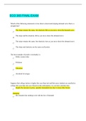
Lateralization: symmetry and asymmetry
Many functions are the same across inter-hemispheres homologues
- primary perception areas with cross-lateralized fields: vision, touch
- Sensory and motor homunculus (at the right)
- Auditory perception is partly bilateral!
Functional asymmetries
- Language usually left-lateralized, nonverbal material on the right
- Global perception is usually right-lateralized, local perception on the left
- Creative vs logical contrast does not hold, most functions need both
hemispheres to be performed correctly
→ Think about split brains studies!
Gyri & Sulci: the wrinkled surface of the cortex
- Gyrus: bump or ridge in the wrinkles
- Sulcus: groove in the wrinkles, some are also called fissures (really big sulcus)
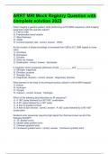
Ventricles of the brain
Open spaces in brain images
- Lateral ventricles
- III rd ventricles
- Aquaduct
- IV th ventricle
- Central canal
Lecture 1 – The brain and cognition over the life spam – 5/9/22
Brain structure & anatomy
Various types of neural and glia cells classified based on their shape, location and function:
Neurons: different cell types categorized by shape and function
- Sensory (afferent = towards the brain)
- Interneurons (stellate, pyramidal, pukinje)
- Motor (efferent = away from the brain) – sending out motor functions
Neurons communicate through action potentials and synaptic transfer
Action potentials – sending these signals
- Thresholded, non-decremental, all-or-nothing response
- What can vary? Rhytm, how many potentials are being fired off
- Triggered by summation of excitatory potentials
- Driven by varying ion permeability of cell membrane
- Propagates along axon (can travel for a meter or more)
- Triggers neurotransmitter release at axon terminal
Synapse:
Action potentials leads to neurotransmitter release into synaptic cleft:
- Some neurons can release more than one type of NT, depending on type of stimulation (low vs. high
frequency stimulation): Acetylcholine, dopamine, norepinephrine, serotonin, glutamate, GABA (few)
- Receptor cells in the postsynaptic membrane can adapt to under- or overuse
- The distribution of synapses connecting to a cell influences its excitability (summation, whether an
axtion potential fires or not)
Glia cells
Vary in shape, size and function
- Astrocytes: help with structural support and blood-brain barrier
- Oligodendrocytes: myelin (allows action potential to go faster) for CNS (central nervous system)
neurons
- Microglial cells: fight infections, waste disposal
,- Ependymal cells: ventricular surface epithelium, create CSF (carinal spinal flued)
- Schwann cells: myelin for peripheral neurons (so outside of the brain)
Grey matter consists of neuronal cell bodies and glia cells, situated on the outer layer of the
hemisphere (cortex) and in subcortical nuclei
Cortical cell layers (cortex – outside layers of the hemisphere)
- Different type of neuron (cells) are often organized in layers
- Sensory (input), interneurons (relay) and motor (output) neurons are grouped
- Layers are different in different crotical areas, depending on primary function
White matter consists of neuronal axons and glia cells, organized in bundles
that connects different grey matter areas
White matter tracs – bundles of myelinated axons (myelin looks white)
Connecting neurons throughout the central and peripheral nervous system
- Association fibers connecting areas within a hemisphere (same side)
- Commissural fibers crossing to the other hemisphere,
to the same (homotopic) or a different place (heterotopic)
- Projection fibers connect outwards, to subcortical regions, cerebellum
or the spinal chord
→ different types of connections – sending signals outwards or in brain
Main components of the brain can be identified and subdivided into specific areas:
Major components of the CNS
- Forebrains: incl hemispheres, corpus callosum and subcortical deep structures (telencephalon)
- Diencephalon: incl thalamic structures
- Midbrain (mesencephalon) - top of brain stem; incl motor and sensory relay nuclei
- Hindbrain (metencephalon); incl pons and cerebellum, medulla oblongata
- Spinal chord
Hindbrain and midbrain:
Hindbrain:
- Medulla oblongata: contributes to vital reflexes, damage is often fatal
- Pons: Crossing over of many fibers in the motor and sensory pathways for contralateral motor
control
- Cerebellum: automated movement, balance, timing & time perfection, sensorimotor coupling,
attention shifting, other cognitive functions
- Origin of cranial nerves V-XII:
Midbrain/ mesecephalon:
- Superior/ inferior colliculi: contribute to sensory processing
- Substantia nigra: contributes to movement initiation
- Origin of cranial nerves II-IV
,Diencephalon:
- Thalamus: important relay station (many subnuclei); any information that goes in or out passes
through the thalamus at some point → delivers signals to almost the whole brain
- Hypothalamus, pituary gland
Some areas are highly specialized, and some areas are just a relay station/ gating functions
Thalamus:
Telencephalon (or cerebrum): subcortical areas: anything underneath the cortex
- Thalamus
- Basal ganglia (see underneath): located deep in the brain
- Limbic structures
Basal ganglia circuits:
Multiple circuits in the brain go through the basal ganglia:
- Motor circuit: organizing voluntary movement through inhibitory excitatory pathways
- Associative ciruit: contributing to learning, predictive processing, sequencing (sequence of
movements/ learning)
- Reward circuit: producing pleasure responses, motivational functions
→ circuits have functionally relation to each other: reward is important for learning etc
Limbic system (stress response, emotional processing):
- Cingulate (part of cortex, so not entirely subcortical)
- Hippocampus
- Hypothalamus
- Amygdala
→ emotional coloring of what you experience
, Telencephalon: cortical lobes (not the cerebellum):
- Frontal: EF, planning, impulse control, motor function, reward circuit, short-term memory etc
→ mainly about output, behavior, how we manage ourself in the world
- Parietal: sensory integration, association processes, language functions, spatial processing, sense of
touch, some visual processes etc
- Occipital: mainly primary visual areas (lot of aspects of sight)
- Temporal: memory, emotion association, primary auditory areas (music!), some visual processes etc
Brain areas are not necessarily mapped one-to-one with specific functions
Although many brain areas have homologues in each hemisphere,
some areas do not!
Lateralization: symmetry and asymmetry
Many functions are the same across inter-hemispheres homologues
- primary perception areas with cross-lateralized fields: vision, touch
- Sensory and motor homunculus (at the right)
- Auditory perception is partly bilateral!
Functional asymmetries
- Language usually left-lateralized, nonverbal material on the right
- Global perception is usually right-lateralized, local perception on the left
- Creative vs logical contrast does not hold, most functions need both
hemispheres to be performed correctly
→ Think about split brains studies!
Gyri & Sulci: the wrinkled surface of the cortex
- Gyrus: bump or ridge in the wrinkles
- Sulcus: groove in the wrinkles, some are also called fissures (really big sulcus)
Ventricles of the brain
Open spaces in brain images
- Lateral ventricles
- III rd ventricles
- Aquaduct
- IV th ventricle
- Central canal