Lectures course 2.5: Use it or Lose it
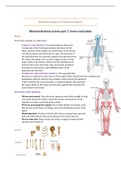
Musculoskeletal system part 1: bones and joints
Bones
Your body contains c.a. 200 bones
- Central/ axial skeleton: The axial skeleton forms the
central axis of the body and includes the bones of the
skull, ossicles of the middle ear, hyoid bone of the throat,
vertebral column, and the thoracic cage. The function of
the axial skeleton is to provide support and protection for
the brain, the spinal cord, and the organs in the ventral
body cavity. It provides a surface for the attachment of
muscles that move the head, neck, and trunk, performs
respiratory movements, and stabilizes parts of the
appendicular skeleton.
- Peripheral/ appendicular skeleton: The appendicular
skeleton is composed of the bones of the upper limbs (which function to grasp and
manipulate objects) and the lower limbs (which permit locomotion).
It also includes the pectoral girdle, or shoulder girdle, that attaches
the upper limbs to the body, and the pelvic girdle that attaches the
lower limbs to the body
Functions of the skeleton:
- Allows movement: Your skeleton supports your body weight to help
you stand and move. Joints, connective tissue and muscles work
together to make your body parts mobile
- Protects and supports organs: Your skull shields your brain, your
ribs protect your heart and lungs, and your backbone protects your
spine
- Hematopoiesis: Produces blood cells: Bones contain bone marrow.
Red and white blood cells are produced in the bone marrow
- Stores minerals: Bones hold your body’s supply of minerals like
calcium and vitamin D.
,Parts the skeletal system:
- Periosteum: The periosteum is a tough membrane that covers and protects the outside of
the bone.
- Compact bone: Below the periosteum, compact bone is white, hard, and smooth. It
provides structural support and protection.
- Spongy bone: The core, inner layer of the bone is softer than compact bone. It has small
holes called pores to store marrow.
- Cartilage: This smooth and flexible substance covers the tips of your bones where they
meet. It enables bones to move without friction (rubbing against each other).
- Joints: A joint is where two or more bones in the body come together
- Ligaments: Bands of strong connective tissue called ligaments hold bones together.
- Tendons: Tendons are bands of tissue that connect the ends of a muscle to your bone
Development of long bone growth:
Initial Bone Formation
The formation of bone during the fetal stage of development occurs by two processes:
intramembranous ossification and endochondral ossification.
Intramembranous Ossification
Intramembranous ossification mainly occurs during the formation of the flat bones of the skull,
as well as the mandible, maxilla, and clavicles. The bone is formed from connective tissue such as
mesenchyme tissue rather than from cartilage. The steps in intramembranous ossification are:
- Development of ossification center
- Calcification
- Formation of trabeculae
- Development of periosteum
Endochondral Ossification
Endochondral ossification begins with points in the cartilage called “primary ossification
centers.” They mostly appear during fetal development, though a few short bones begin their
,primary ossification after birth. These cartilage points are responsible for the formation of the
diaphysis of long bones, short bones, and certain parts of irregular bones.
Secondary ossification occurs after birth and forms the epiphyses of long bones and the
extremities of irregular and flat bones. The diaphysis and both epiphyses of a long bone are
separated by a growing zone of cartilage (the epiphyseal plate). When the child reaches skeletal
maturity (18 to 25 years of age), all cartilage is replaced by bone, fusing the diaphysis and both
epiphyses together (epiphyseal closure).
Remodeling
Remodeling or bone turnover is the process of resorption followed by replacement of bone with
little change in shape, and occurs throughout a person’s life, long beyond the initial development
of bone. Osteoblasts and osteoclasts, coupled together via paracrine cell signaling, are referred
to as a bone remodeling unit. Approximately 10% of the skeletal mass of an adult is remodeled
each year.
The purpose of remodeling is to regulate calcium homeostasis and repair micro-damage from
everyday stress, as well as to shape the skeleton during growth. Repeated stress, such as weight-
bearing exercise or bone healing, results in the bone thickening at the points of maximum stress
(Wolff’s law).
Bone grows in length because cartilage grows and cartilage is replaced by bone. Bone tissue is
active tissue: during growth bones also remodels, also after fracture and homeostasis
Continuously simultaneous process of bone deposit (osteoblasts) and bone resorption
(osteoclasts) this is regulated by hormones like PTH and calcitonin. Mechanical loading is also
important
The way we use our bones play an important role in how they develop:
- Wolff's Law states that your bones will adapt based on the stress or demands placed on
them. When you work your muscles, they put stress on your bones. In response, your
bone tissue remodels and becomes stronger. ... If you don't use the muscles surrounding
a bone much, the bone tissue can weaken
- when there is weight loaded at a bone, it results in a bow, wherein there is compression
of the inside and tension of the outside of the bow
- mechanical loading comes from gravity, body weight and muscle force
Reinforcements (ridges, protrusions) develop on sites where muscles attach. These
reinforcements develop during life. Young babies who did not have a great exposure to gravity
and load yet
Joints
A joint is where two or more bones in the body come together and they ensure mobilization
Structural classification:
- Fibrous joints: Fibrous joints are connected by dense connective tissue consisting mainly
of collagen. These joints are also called fixed or immovable joints because they do not
move, like the joints between your skull bones. Fibrous joints have no joint cavity and
are connected via fibrous connective tissue.
- Cartilaginous joints: cartilaginous joints are connected entirely by cartilage
(fibrocartilage or hyaline). Cartilaginous joints allow more movement between bones
, than a fibrous joint but less than the highly mobile synovial joint. The joint between the
manubrium and the sternum is an example of a cartilaginous joint. This type of joint also
forms the growth regions of immature long bones and the intervertebral discs of the
spinal column. The joints in your rib cage are partly movable joints.
- Synovial joints: A synovial joint, also known as a diarthrosis, is the most common and
most movable type of joint in a mammal’s body. Diarthroses are freely movable
articulations. In these joints, the contiguous bony surfaces are covered with articular
cartilage and connected by ligaments lined by synovial membrane. The joint may be
divided, completely or incompletely, by an articular disk or meniscus, the periphery of
which is continuous with the fibrous capsule while its free surfaces are covered by
synovial membrane.
Musculoskeletal system part 2: muscles and joints
Muscles
We have c.a. 600 muscles in our body
Functions muscular system:
- Maintain posture
- Produce movement
- Generate heat
Relations:
- Attachment to skeleton
- Control by nervous system
- Nutrition by vascular system
Muscle shapes:
- Circular Muscles: These muscles appear circular in
shape and are normally sphincter muscles which
surround an opening such as the mouth, surrounded
by Obicularis Oris and Obicularis Oculi surrounding
the eyes
- Parallel muscles: have fibers which, run parallel to each other and are sometimes called
strap muscles. They are normally long muscles which cause large movements, are not
very strong but have good endurance.