1.2 Revision notes
ULTRASTRUCTURE OF CELLS
Electron versus light microscopes
Electron microscopes have a higher resolution than light microscopes
Transmission electron microscopes (TEM): generate high resolution cross-section of objects
Scanning electron microscopes (SEM): enhanced death to map surface of objects in 3D
Resolution: ability of a microscope to making separate two close object
Light microscope Electron microscope
Resolution 0.2 μm (2 nanometres) 0.0001 μm (1 nanometres) –
can provide clearer and
detailed structures
Wavelength Longer (400 – 700nm) Shorter wavelength
Magnification x400 Greater magnification – can
detect smaller resolutions
Prokaryotic cells
‘pro – before’ ‘karyon - nucleus’
Organisms that lack a nucleus
Cytoplasm is not divided into compartments, simple structure
Belong to kingdom Monera
Domains:
- Archaebacteria: found in extreme conditions (temperatures, pH)
- Eubacteria: bacteria in pathogenic form
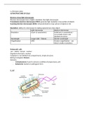
E. coli
, Cytoplasm: fluid component of cell where metabolic reaction occurs, contain ribosomes
Nucleoid: region of cytoplasm where DNA is located (circular), lighter in colour
Plasmids: autonomous circular DNA that can be transferred between prokaryotic cells
(horizontal gene transfer)
70S Ribosomes: RNA and proteins where polypeptide synthesis occurs
Cell membrane: semi permeable and selective barrier around the cell
Cell wall: contains peptidoglycan. Maintains shape and prevents bursting
Slime capsule: polysaccharide layer that prevents cell from drying out and protection from
phagocytosis.
Flagellum: tail that moves cell
Pili: hair-like extensions that enable adherence to surfaces (attachment pili) or bacterial
conjugation (sex pili)
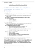
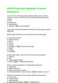
Prokaryotic cell division
Binary fission
- Asexual reproduction
- Single circular chromosome is replicated in response to a replication signal
- Two copies of the chromosome move to opposite sides of the cell and attach to
membrane
- Division of the cell via cytokinesis (membrane elongates and pinches off)
- Each daughter cell contains one copy of chromosome
ULTRASTRUCTURE OF CELLS
Electron versus light microscopes
Electron microscopes have a higher resolution than light microscopes
Transmission electron microscopes (TEM): generate high resolution cross-section of objects
Scanning electron microscopes (SEM): enhanced death to map surface of objects in 3D
Resolution: ability of a microscope to making separate two close object
Light microscope Electron microscope
Resolution 0.2 μm (2 nanometres) 0.0001 μm (1 nanometres) –
can provide clearer and
detailed structures
Wavelength Longer (400 – 700nm) Shorter wavelength
Magnification x400 Greater magnification – can
detect smaller resolutions
Prokaryotic cells
‘pro – before’ ‘karyon - nucleus’
Organisms that lack a nucleus
Cytoplasm is not divided into compartments, simple structure
Belong to kingdom Monera
Domains:
- Archaebacteria: found in extreme conditions (temperatures, pH)
- Eubacteria: bacteria in pathogenic form
E. coli
, Cytoplasm: fluid component of cell where metabolic reaction occurs, contain ribosomes
Nucleoid: region of cytoplasm where DNA is located (circular), lighter in colour
Plasmids: autonomous circular DNA that can be transferred between prokaryotic cells
(horizontal gene transfer)
70S Ribosomes: RNA and proteins where polypeptide synthesis occurs
Cell membrane: semi permeable and selective barrier around the cell
Cell wall: contains peptidoglycan. Maintains shape and prevents bursting
Slime capsule: polysaccharide layer that prevents cell from drying out and protection from
phagocytosis.
Flagellum: tail that moves cell
Pili: hair-like extensions that enable adherence to surfaces (attachment pili) or bacterial
conjugation (sex pili)
Prokaryotic cell division
Binary fission
- Asexual reproduction
- Single circular chromosome is replicated in response to a replication signal
- Two copies of the chromosome move to opposite sides of the cell and attach to
membrane
- Division of the cell via cytokinesis (membrane elongates and pinches off)
- Each daughter cell contains one copy of chromosome