
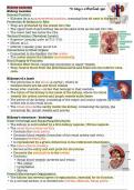
Kidney anatomy
Kidney location
General Position
• Kidneys lie in a paravertebral position, meaning they sit next to the spine.
Protection & Relation to Ribs
• They are protected by the lowest two ribs.
• The upper half of each kidney lies at the same level as the last two ribs.
• The lower half lies below the ribs.
Vertical Position (Vertebral Levels)
• Superior (cranial) pole: at T11–T12.
• Hilum: at L1.
• Inferior (caudal) pole: at L2–L3.
Connections to Other Organs
• Connected to the bladder via the ureter.
• The ureter enters the bladder dorsolaterally.
Blood Supply & Function
• Kidneys filter blood, requiring connection to major vessels.
• They receive blood from the abdominal aorta and drain into the inferior vena
cava.
Kidneys of a lamb
• The pelvis lies in the sinus (a cavity), where in
between there are fat and blood vessels
• Renal lobe= medulla + cortex that belongs to that medulla
• The hilum of the kidney is the navel of the kidney, where the blood
vessels the ureter, nerves and lymph vessels enter/leave.
• The pelvis of the kidney, consisting of the major and minor calyces up to the
ureter sits in the renal sinus.
• The renal sinus is the cavity inside the kidney, containing the pelvis, the
intrarenal fat, lymph vessels and nerves.
Kidney’s structure - histology
Outer Coverings and Supporting Structures
• The kidney is surrounded by a firm kidney capsule / fibrous capsule.
• The renal sinus (renal cavity):
• Contains the renal pelvis.
• Contains blood vessels (branches of the renal artery and vein).
• Contains perirenal fat.
• Is represented by a green dotted line in the referenced illustration.
• The hilum (hilum renale):
• Serves as the entry and exit point for structures.
• Connects the renal pelvis to the ureter.
• Contains:
• Renal blood vessels (arteries and veins)
• The ureter
• Lymphatics
• Nerves
Overall Macroscopic Organization
• The kidney has several layers of organization, essential for its function.
• In a coronal section, it is divided into: Cortex, Medulla, Renal pelvis
,Cortex
• The cortex contains the structures where the initial blood filtration occurs.
• Contains renal corpuscles (Bodies of Malpighi):
• Not visible to the naked eye.
• Each renal corpuscle includes:
• A glomerulus (capillary network)
• A Bowman’s capsule
• Renal corpuscles are only found in the cortex, not in the medulla.
• Primary urine (≈180 L/day) is produced here by filtration.
• Cortex extends between medullary pyramids as renal columns (columns of
Bertini).
• Medullary rays:
• Bundles of parallel renal tubules from the medulla that penetrate the cortex.
• Located at the center of each lobule.
• Appear “a bit like sunrays”.
• Structures mainly found in a lobule:
• Renal corpuscles
• Medullary rays (collecting ducts)
• Afferent and efferent arterioles
• Renal tubules
Medulla
• The medulla consists of 10–18 pyramids (sometimes described as ≈10–15
macroscopically).
• The medulla is responsible for concentrating the primary urine.
• Kidney tubules present in the medulla perform urine concentration:
• Not visible macroscopically.
• Degree of urine concentration depends on fluid and salt intake.
• Pyramids are separated by renal columns.
• Predominant histological structures in the medulla: Collecting ducts
Renal Lobes and Lobules
Renal Lobe
• A renal lobe consists of:
• One medullary pyramid
• Its associated cortex.
• From the histology text:
• “One pyramid with its concomitant cortex forms a renal lobe.”
Lobules
• Each lobe is subdivided into lobules.
• Each lobule lies between two interlobular arteries.
• Each lobule contains:
• Numerous nephrons, the filtering units.
Nephrons and Filtration
• A nephron consists of: a renal corpuscle and a renal tubule
• Blood is filtered in the corpuscle, producing primary urine.
• Total primary urine production: ≈ 180 L/day.
• Filtrate (primary urine):
• Is concentrated in the tubules.
• Is eventually excreted at the top of each pyramid, the renal papilla.
• The papilla has a cribriform area through which urine passes.
,Urine Flow Pathway
1. Renal papilla (tip of pyramid)
2. Minor calyx (calyx minor)
• Urine drips into it through pores at the papilla
(cribriform area).
3. Major calyx (calyx major)
• Formed by 4–5 minor calyces.
4. Renal pelvis (pelvis renalis)
• Formed by 2–3 major calyces.
• Fills most of the renal sinus.
5. Ureter
• The renal pelvis connects to the ureter at the hilum.
Kidney Segments (Surgical & Vascular Anatomy)
• Each kidney has 4–5 segments, each with its own blood supply.
• Each segment is supplied by a segmental artery.
• There are no segmental veins.
• Surgical principle:
• If a structure within one segment must be removed, the whole segment must
be removed.
• Partial removal results in death of the remaining part of that segment (due to
loss of independent blood supply).
Blood Vessel Organization
• Major vessels:
• Renal arteries and renal veins
• There may be more than one renal artery per kidney.
• Segmental arteries (one per kidney segment)
• Interlobar arteries and veins
• Arcuate arteries and veins
• Interlobular arteries and veins
• Also called cortical radiate arteries and veins
Renal vascularisation
• The renal blood vessels are critically important because the kidney’s major
functions—filtration of blood and excretion of excess fluid and salt as urine—
depend on precise and organized vascular pathways.
• The renal artery (arteria renalis) enters the kidney via the hilum.
• The kidney also has a renal vein (vena renalis), which ultimately returns
blood to systemic circulation.
• After entering at the hilum, the renal artery splits into segmental arteries
(arteriae segmentales).
• Typically 5 segmental arteries arise just before or just after the renal artery
enters the hilum.
• Each segmental artery supplies one kidney segment, and each kidney
segment consists of several lobes.
• Segmental arteries may give blood to adjacent lobes or lobes situated behind
one another.
• There are no segmental veins (even tho some diagrams sometimes show them).
• Segmental arteries branch into interlobar arteries (arteriae interlobares).
• These run between the lobes (i.e., between the medullary pyramids), exactly
as their name suggests.
• Interlobar veins run parallel to these arteries.
, • At the border between cortex and medulla, interlobar arteries branch into
arcuate arteries (arteriae arcuatae).
• “Arcuate” (arcus = arch/bow) describes their arch-shaped course along the
corticomedullary junction.
• Arcuate veins run parallel to these arteries.
• Arcuate arteries give rise to interlobular arteries (arteriae interlobulares), also
known as cortical radiate arteries.
• These arteries lie between the lobules, and the cortex is composed of many
lobules separated by these vessels.
• Interlobular veins run parallel to these arteries.
• From each interlobular (cortical radiate) artery, several afferent arterioles
originate.
• Afferent arterioles carry blood into the glomeruli of the renal corpuscles.
• Afferent arterioles → Glomerulus → filtration occurs.
• After filtration in the glomerulus, blood exits via efferent arterioles.
• Efferent arterioles then branch into a capillary network, followed by venules
and ultimately veins.
• Efferent arterioles → leave the renal corpuscle → supply capillary beds before
draining into venous vessels.
• Structures Mainly Found Within a Lobule
• Renal corpuscles
• Medullary rays (collecting ducts, darker in histology)
• Afferent and efferent arterioles
• Renal tubules
• Capillaries
• Full vascularization pathway
Renal artery → Segmental arteries → Interlobar arteries → Arcuate arteries →
Interlobular (cortical radiate) arteries → Afferent arterioles → Glomerular
capillaries → Efferent arterioles → Capillary networks → Interlobular (cortical
radiate) veins → Arcuate veins → Interlobar veins → Renal vein
• Anatomical Regions
• The arterial pathway runs through:
• Medullary pyramids / pyramids of the kidney
• Cortex
• Interlobar arteries are located between pyramids/lobes.
• Interlobular arteries are located between lobules.
Kidney location
General Position
• Kidneys lie in a paravertebral position, meaning they sit next to the spine.
Protection & Relation to Ribs
• They are protected by the lowest two ribs.
• The upper half of each kidney lies at the same level as the last two ribs.
• The lower half lies below the ribs.
Vertical Position (Vertebral Levels)
• Superior (cranial) pole: at T11–T12.
• Hilum: at L1.
• Inferior (caudal) pole: at L2–L3.
Connections to Other Organs
• Connected to the bladder via the ureter.
• The ureter enters the bladder dorsolaterally.
Blood Supply & Function
• Kidneys filter blood, requiring connection to major vessels.
• They receive blood from the abdominal aorta and drain into the inferior vena
cava.
Kidneys of a lamb
• The pelvis lies in the sinus (a cavity), where in
between there are fat and blood vessels
• Renal lobe= medulla + cortex that belongs to that medulla
• The hilum of the kidney is the navel of the kidney, where the blood
vessels the ureter, nerves and lymph vessels enter/leave.
• The pelvis of the kidney, consisting of the major and minor calyces up to the
ureter sits in the renal sinus.
• The renal sinus is the cavity inside the kidney, containing the pelvis, the
intrarenal fat, lymph vessels and nerves.
Kidney’s structure - histology
Outer Coverings and Supporting Structures
• The kidney is surrounded by a firm kidney capsule / fibrous capsule.
• The renal sinus (renal cavity):
• Contains the renal pelvis.
• Contains blood vessels (branches of the renal artery and vein).
• Contains perirenal fat.
• Is represented by a green dotted line in the referenced illustration.
• The hilum (hilum renale):
• Serves as the entry and exit point for structures.
• Connects the renal pelvis to the ureter.
• Contains:
• Renal blood vessels (arteries and veins)
• The ureter
• Lymphatics
• Nerves
Overall Macroscopic Organization
• The kidney has several layers of organization, essential for its function.
• In a coronal section, it is divided into: Cortex, Medulla, Renal pelvis
,Cortex
• The cortex contains the structures where the initial blood filtration occurs.
• Contains renal corpuscles (Bodies of Malpighi):
• Not visible to the naked eye.
• Each renal corpuscle includes:
• A glomerulus (capillary network)
• A Bowman’s capsule
• Renal corpuscles are only found in the cortex, not in the medulla.
• Primary urine (≈180 L/day) is produced here by filtration.
• Cortex extends between medullary pyramids as renal columns (columns of
Bertini).
• Medullary rays:
• Bundles of parallel renal tubules from the medulla that penetrate the cortex.
• Located at the center of each lobule.
• Appear “a bit like sunrays”.
• Structures mainly found in a lobule:
• Renal corpuscles
• Medullary rays (collecting ducts)
• Afferent and efferent arterioles
• Renal tubules
Medulla
• The medulla consists of 10–18 pyramids (sometimes described as ≈10–15
macroscopically).
• The medulla is responsible for concentrating the primary urine.
• Kidney tubules present in the medulla perform urine concentration:
• Not visible macroscopically.
• Degree of urine concentration depends on fluid and salt intake.
• Pyramids are separated by renal columns.
• Predominant histological structures in the medulla: Collecting ducts
Renal Lobes and Lobules
Renal Lobe
• A renal lobe consists of:
• One medullary pyramid
• Its associated cortex.
• From the histology text:
• “One pyramid with its concomitant cortex forms a renal lobe.”
Lobules
• Each lobe is subdivided into lobules.
• Each lobule lies between two interlobular arteries.
• Each lobule contains:
• Numerous nephrons, the filtering units.
Nephrons and Filtration
• A nephron consists of: a renal corpuscle and a renal tubule
• Blood is filtered in the corpuscle, producing primary urine.
• Total primary urine production: ≈ 180 L/day.
• Filtrate (primary urine):
• Is concentrated in the tubules.
• Is eventually excreted at the top of each pyramid, the renal papilla.
• The papilla has a cribriform area through which urine passes.
,Urine Flow Pathway
1. Renal papilla (tip of pyramid)
2. Minor calyx (calyx minor)
• Urine drips into it through pores at the papilla
(cribriform area).
3. Major calyx (calyx major)
• Formed by 4–5 minor calyces.
4. Renal pelvis (pelvis renalis)
• Formed by 2–3 major calyces.
• Fills most of the renal sinus.
5. Ureter
• The renal pelvis connects to the ureter at the hilum.
Kidney Segments (Surgical & Vascular Anatomy)
• Each kidney has 4–5 segments, each with its own blood supply.
• Each segment is supplied by a segmental artery.
• There are no segmental veins.
• Surgical principle:
• If a structure within one segment must be removed, the whole segment must
be removed.
• Partial removal results in death of the remaining part of that segment (due to
loss of independent blood supply).
Blood Vessel Organization
• Major vessels:
• Renal arteries and renal veins
• There may be more than one renal artery per kidney.
• Segmental arteries (one per kidney segment)
• Interlobar arteries and veins
• Arcuate arteries and veins
• Interlobular arteries and veins
• Also called cortical radiate arteries and veins
Renal vascularisation
• The renal blood vessels are critically important because the kidney’s major
functions—filtration of blood and excretion of excess fluid and salt as urine—
depend on precise and organized vascular pathways.
• The renal artery (arteria renalis) enters the kidney via the hilum.
• The kidney also has a renal vein (vena renalis), which ultimately returns
blood to systemic circulation.
• After entering at the hilum, the renal artery splits into segmental arteries
(arteriae segmentales).
• Typically 5 segmental arteries arise just before or just after the renal artery
enters the hilum.
• Each segmental artery supplies one kidney segment, and each kidney
segment consists of several lobes.
• Segmental arteries may give blood to adjacent lobes or lobes situated behind
one another.
• There are no segmental veins (even tho some diagrams sometimes show them).
• Segmental arteries branch into interlobar arteries (arteriae interlobares).
• These run between the lobes (i.e., between the medullary pyramids), exactly
as their name suggests.
• Interlobar veins run parallel to these arteries.
, • At the border between cortex and medulla, interlobar arteries branch into
arcuate arteries (arteriae arcuatae).
• “Arcuate” (arcus = arch/bow) describes their arch-shaped course along the
corticomedullary junction.
• Arcuate veins run parallel to these arteries.
• Arcuate arteries give rise to interlobular arteries (arteriae interlobulares), also
known as cortical radiate arteries.
• These arteries lie between the lobules, and the cortex is composed of many
lobules separated by these vessels.
• Interlobular veins run parallel to these arteries.
• From each interlobular (cortical radiate) artery, several afferent arterioles
originate.
• Afferent arterioles carry blood into the glomeruli of the renal corpuscles.
• Afferent arterioles → Glomerulus → filtration occurs.
• After filtration in the glomerulus, blood exits via efferent arterioles.
• Efferent arterioles then branch into a capillary network, followed by venules
and ultimately veins.
• Efferent arterioles → leave the renal corpuscle → supply capillary beds before
draining into venous vessels.
• Structures Mainly Found Within a Lobule
• Renal corpuscles
• Medullary rays (collecting ducts, darker in histology)
• Afferent and efferent arterioles
• Renal tubules
• Capillaries
• Full vascularization pathway
Renal artery → Segmental arteries → Interlobar arteries → Arcuate arteries →
Interlobular (cortical radiate) arteries → Afferent arterioles → Glomerular
capillaries → Efferent arterioles → Capillary networks → Interlobular (cortical
radiate) veins → Arcuate veins → Interlobar veins → Renal vein
• Anatomical Regions
• The arterial pathway runs through:
• Medullary pyramids / pyramids of the kidney
• Cortex
• Interlobar arteries are located between pyramids/lobes.
• Interlobular arteries are located between lobules.



