1 of 10
B2- Basic components of living things
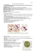
2.1.1 Cell structure
(a) The use of microscopy to observe and investigate different types of cell and cell structure in a
range of eukaryotic organisms.
Key words:
Chromatic aberration- failure of a lens to focus all colours to the same point.
Brightfield microscopy- illumination light is transmitted through the sample and the contrast is
generated by the absorption of light in dense areas of the specimen.
Wide-field microscopy- any technique in which the entire specimen of interest is exposed to the
light source resulting in an image.
Fluoresce- give off light.
Focal plane- the distance that gives the sharpest focus.
Compound light microscope
• Has two lenses, objective (near the specimen) and eyepiece lens (through which the specimen is
viewed).
• The lens configuration allows for a much higher magnification and reduces chromatic aberration
than a simple light microscope.
• Illuminated by white light below sample (brightfield microscopy), or above if opaque.
• The whole sample illuminated at once is known as wide-field microscopy.
• Light first passes through a condenser lens, doesn’t magnify, it focuses light on the sample
being observed.
• The cytosol of cells and other cell structures are often transparent.
• Lower resolution than electron microscope, 0.2μm.
• Used to look at whole cells or tissue.
• Magnification of about x1500.
Electron microscope
• A beam of electrons with a wavelength of less than 1 nanometre is used to highlight the
specimen.
• More detail of cell ultrastructures can be views as electrons have much smaller wavelength than
light waves.
• Able to produce images with magnification of up to
x500,000 and resolution up to 0.0002μm.
Transmission electron microscope (TEM)
• Use electromagnets to focus a beam of electrons, which
is then transmitted through the specimen. Similar to light
microscopy.
• Denser parts of the specimen absorb more electrons,
look darker on image.
• Highest resolution, 0.0002μm, as they can distinguish
between the smallest objects.
, 2 of 10
• Magnification can be more than x1,000,000.
• Resolving power of 0.5nm.
Scanning electron microscope (SEM)
• Scan a beam of electrons across the surface of a
specimen.
• Which knocks off electrons from the specimen, which
are gathered in a cathode ray tube to form an image.
• Image shows surface of the specimen and is 3-D.
• Maximum resolution of 0.002μm and maximum
magnification of usually less than x500,000.
• Resolving power from 3-10nm, lower.
Laser scanning confocal microscope
• Moves a single spot of focused light across a
specimen, which is usually tagged with
fluorescent dye.
• Laser causes the dye to fluoresce, which is
then filtered through a pinhole aperture onto a
detector.
• Detector, connected to a computer, generate
an image, can be 3-D.
• Pinhole means any out-of-focus light is
blocked, so they produce a much clearer image
than normal light microscopes.
• Can be use to look at objects at different
depths in thick specimens.
• Non- invasive used in the diagnosis of diseases
of the eye
• Position of the two pinholes means light waves
from the laser follow the same path as the light
waves radiated when the sample fluoresces,
both have the same focal plane, hence
confocal.
(b) The preparation and examination of microscope slides for use in light microscopy.
Key words:
Sectioning- cutting specimens into thin slices with a blade.
Eyepiece graticule- a glass disc marked with a fine scale of 1 to 100.
Stage micrometer- microscope slide with a very accurate scale in μm engraved on it.
Preparation
Dry mount Wet mount Squash slides Smear slides
How to • Solid specimens • Specimens are • After wet mount • Edge of a slide is
cut into thin suspended in a prepared. used to smear a
slices with a liquid. • Lens tissue is sample, creating
blade, sectioning. • Cover slip is used to gently a thin, even
• Specimen placed placed on from press down the coating on
in the centre of an angle. cover slip. another slide.
the slide and a • Damage to cover • Cover slip is then
cover slop is slip can be placed over the
placed over the avoided by sample.
sample. squashing the
sample with two
microscope
slides.
B2- Basic components of living things
2.1.1 Cell structure
(a) The use of microscopy to observe and investigate different types of cell and cell structure in a
range of eukaryotic organisms.
Key words:
Chromatic aberration- failure of a lens to focus all colours to the same point.
Brightfield microscopy- illumination light is transmitted through the sample and the contrast is
generated by the absorption of light in dense areas of the specimen.
Wide-field microscopy- any technique in which the entire specimen of interest is exposed to the
light source resulting in an image.
Fluoresce- give off light.
Focal plane- the distance that gives the sharpest focus.
Compound light microscope
• Has two lenses, objective (near the specimen) and eyepiece lens (through which the specimen is
viewed).
• The lens configuration allows for a much higher magnification and reduces chromatic aberration
than a simple light microscope.
• Illuminated by white light below sample (brightfield microscopy), or above if opaque.
• The whole sample illuminated at once is known as wide-field microscopy.
• Light first passes through a condenser lens, doesn’t magnify, it focuses light on the sample
being observed.
• The cytosol of cells and other cell structures are often transparent.
• Lower resolution than electron microscope, 0.2μm.
• Used to look at whole cells or tissue.
• Magnification of about x1500.
Electron microscope
• A beam of electrons with a wavelength of less than 1 nanometre is used to highlight the
specimen.
• More detail of cell ultrastructures can be views as electrons have much smaller wavelength than
light waves.
• Able to produce images with magnification of up to
x500,000 and resolution up to 0.0002μm.
Transmission electron microscope (TEM)
• Use electromagnets to focus a beam of electrons, which
is then transmitted through the specimen. Similar to light
microscopy.
• Denser parts of the specimen absorb more electrons,
look darker on image.
• Highest resolution, 0.0002μm, as they can distinguish
between the smallest objects.
, 2 of 10
• Magnification can be more than x1,000,000.
• Resolving power of 0.5nm.
Scanning electron microscope (SEM)
• Scan a beam of electrons across the surface of a
specimen.
• Which knocks off electrons from the specimen, which
are gathered in a cathode ray tube to form an image.
• Image shows surface of the specimen and is 3-D.
• Maximum resolution of 0.002μm and maximum
magnification of usually less than x500,000.
• Resolving power from 3-10nm, lower.
Laser scanning confocal microscope
• Moves a single spot of focused light across a
specimen, which is usually tagged with
fluorescent dye.
• Laser causes the dye to fluoresce, which is
then filtered through a pinhole aperture onto a
detector.
• Detector, connected to a computer, generate
an image, can be 3-D.
• Pinhole means any out-of-focus light is
blocked, so they produce a much clearer image
than normal light microscopes.
• Can be use to look at objects at different
depths in thick specimens.
• Non- invasive used in the diagnosis of diseases
of the eye
• Position of the two pinholes means light waves
from the laser follow the same path as the light
waves radiated when the sample fluoresces,
both have the same focal plane, hence
confocal.
(b) The preparation and examination of microscope slides for use in light microscopy.
Key words:
Sectioning- cutting specimens into thin slices with a blade.
Eyepiece graticule- a glass disc marked with a fine scale of 1 to 100.
Stage micrometer- microscope slide with a very accurate scale in μm engraved on it.
Preparation
Dry mount Wet mount Squash slides Smear slides
How to • Solid specimens • Specimens are • After wet mount • Edge of a slide is
cut into thin suspended in a prepared. used to smear a
slices with a liquid. • Lens tissue is sample, creating
blade, sectioning. • Cover slip is used to gently a thin, even
• Specimen placed placed on from press down the coating on
in the centre of an angle. cover slip. another slide.
the slide and a • Damage to cover • Cover slip is then
cover slop is slip can be placed over the
placed over the avoided by sample.
sample. squashing the
sample with two
microscope
slides.