

Skill- HEENT Demonstrated (check off)
Massage scalp around in circles to Head exam, then I move to your eyes, ears, nose,
palpate and throat.
Head exam
Inspect: Look for your hair distribution, your
head contour, and depression.
Look into your face for any involuntary
movement, scarring, or lesion. Eyebrow, eyelash
should be symmetrical.
Palpate: for any lump, bump, or tender mass.
Feel your occipital.
Feel your temporal pulse/artery.
Open and close your mouth. I can feel your
trigeminal nerve.
Inspect the head and face for symmetry
and deformity, Assess skin
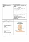
Palpate lymph nodes
Occipital
posterior auricular/pre-auricular,
tonsillar
Submandibular
submental
anterior cervical/ posterior
cervical
supraclavicular/Infraclavicular
Mobile, and non-tender =>Normal
Tender, fix, hard => Abnormal
,Check Sinuses
Frontal sinus
Palpate maxillary sinus
Ethmoid sinus.
**Must name sinuses during
validation**
Inspect the eyes CN II: Optic nerve
assess six positions of gaze CN III: EOM
extraocular movement EOM CN IV: Inward
visual fields CN VI: Outward (Abduction)
ophthalmoscopic evaluation Snellen Chart
including red light reflex Visual Field
pupillary response to light and PERRLA
accommodation Red Reflex
Inspect the eyes: Pull the conjunctiva down.
Should be pink and white. Any foreign body. No
jaundice.
Ostoscope:
+ Check for PERRLA. Direct and consensual
light reaction
+Check visual field: Peripheral vision field.
Confrontational testing. Close one eye and move
your hand inward. Superior and posterior view.
+6 cardinal view of gaze: EOM testing (CN III)
+Move the finger toward her nose : Look for
conversion (Eye toward nose).
+ Look at pupil constrict (Near test). Look at my
finger and back wall (Constrict close object and
dilate at far object)
, Ophthalmoscope exam:
+Red light reflex
The term ‘red reflex’ refers to the reddish-
orange glow or the reflection of light that
is observed from the eye’s retina during
direct ophthalmoscopy.
Healthy eyes where there is no obstruction
in the optical pathway, red reflex is present
Absence of red reflex is indicative of
certain serious eye conditions.
+ 15-degree L hand on pt’s head. Look for red
reflex , follow all the way in until close to pt.
Move your head and scope together. U can see
optic disc.
Inspect external and internal ears with Inspection: extra canal, drainage, cerumen,
otoscope deformity, any pain? If pt has drainage , ear pain,
redness. Don’t inspect that ear, don’t put
ostoscope into his ear.
Otoscope: pull Up and Back
Children Down and Back
Look into canal and tympanic membrane
Light reflex
Tympanic membrane should be nice
pearly
Bulging, any redness, pain, drainage, ear
infection
R ear : Cone of light at 5
L ear: Cone of light at 7
Massage scalp around in circles to Head exam, then I move to your eyes, ears, nose,
palpate and throat.
Head exam
Inspect: Look for your hair distribution, your
head contour, and depression.
Look into your face for any involuntary
movement, scarring, or lesion. Eyebrow, eyelash
should be symmetrical.
Palpate: for any lump, bump, or tender mass.
Feel your occipital.
Feel your temporal pulse/artery.
Open and close your mouth. I can feel your
trigeminal nerve.
Inspect the head and face for symmetry
and deformity, Assess skin
Palpate lymph nodes
Occipital
posterior auricular/pre-auricular,
tonsillar
Submandibular
submental
anterior cervical/ posterior
cervical
supraclavicular/Infraclavicular
Mobile, and non-tender =>Normal
Tender, fix, hard => Abnormal
,Check Sinuses
Frontal sinus
Palpate maxillary sinus
Ethmoid sinus.
**Must name sinuses during
validation**
Inspect the eyes CN II: Optic nerve
assess six positions of gaze CN III: EOM
extraocular movement EOM CN IV: Inward
visual fields CN VI: Outward (Abduction)
ophthalmoscopic evaluation Snellen Chart
including red light reflex Visual Field
pupillary response to light and PERRLA
accommodation Red Reflex
Inspect the eyes: Pull the conjunctiva down.
Should be pink and white. Any foreign body. No
jaundice.
Ostoscope:
+ Check for PERRLA. Direct and consensual
light reaction
+Check visual field: Peripheral vision field.
Confrontational testing. Close one eye and move
your hand inward. Superior and posterior view.
+6 cardinal view of gaze: EOM testing (CN III)
+Move the finger toward her nose : Look for
conversion (Eye toward nose).
+ Look at pupil constrict (Near test). Look at my
finger and back wall (Constrict close object and
dilate at far object)
, Ophthalmoscope exam:
+Red light reflex
The term ‘red reflex’ refers to the reddish-
orange glow or the reflection of light that
is observed from the eye’s retina during
direct ophthalmoscopy.
Healthy eyes where there is no obstruction
in the optical pathway, red reflex is present
Absence of red reflex is indicative of
certain serious eye conditions.
+ 15-degree L hand on pt’s head. Look for red
reflex , follow all the way in until close to pt.
Move your head and scope together. U can see
optic disc.
Inspect external and internal ears with Inspection: extra canal, drainage, cerumen,
otoscope deformity, any pain? If pt has drainage , ear pain,
redness. Don’t inspect that ear, don’t put
ostoscope into his ear.
Otoscope: pull Up and Back
Children Down and Back
Look into canal and tympanic membrane
Light reflex
Tympanic membrane should be nice
pearly
Bulging, any redness, pain, drainage, ear
infection
R ear : Cone of light at 5
L ear: Cone of light at 7



