Module 2: Major Histocompatibility Complex and T cell responses
Major histocompatibility complex
MHC:
Responsible for transplantation rejection
Highly polymorphic exact set and make-up differs between individuals
Non-matching MHC is recognized by T cells and killed
All cell, except red blood cells, express MHC-I
T cell recognition of MHC/peptide complexes
Main function of MHC molecules is to bind to antigens derived from pathogens and display them on
the cell surface for recognition by antigen specific T-cells
T lymphocytes receptor (TCR) recognize combination of MHC and pathogen peptide
MHC molecule contains cleft/groove which binds the peptide
o Cleft is polymorphic variation in peptide fragments that can be bound
o Mutation of virus to escape T cell is not beneficial because next host is different
o A peptide can only bind the peptide binding groove of a certain MHC molecules if it
has certain amino acids at key positions anchor residues
Amino acids that are most often found are called dominant anchor residues
Peptide is called an epitope
MHC class I and MHC class II molecules
two main groups of MHC molecules:
Class I
o Single MHC molecule containing peptide binding cleft
o On the cell surface always associated with β2-microglobulin
o Cleft more closed peptide binding more restricted
Class II
o Two chains, an α and β chain, that together form the peptide binding cleft
o The ends of peptides can stick out of the cleft
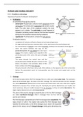
Antigen presentation by MHC-I
Every cell of the body can be infected by a virus or become carcinogenic. These cells will produce
viral or cancer specific (mutated) proteins in the cytosol and peptides of these cytosolic proteins will
be presented on MHC-I molecules on the cell surface
Cytotoxic T cells (CTLs) expressing CD8 molecules on their surface will recognize the MHC-I/peptide
complex and kill the infected or cancerous cell
MHC-I antigen presentation process:
1. Virus in cytoplasm
2. Production of viral proteins in cytosol
3. Viral protein becomes ubiquitinated and are degraded into peptides by proteasome
4. Peptides are transported to ER via transporter associated with antigen processing (TAP)
5. Tapasin brings empty MHC-I to TAP
6. In lumen, peptide is trimmed by ER resident aminopeptidase (ERAP)
7. Peptide and MHC-I form stable complex
8. Complex moves to Golgi complex
9. exocytic vesicles transport peptide-class I complexes to the cell surface
10. complex gets recognized by specific CD8+ T lymphocytes
11. CD8 binds to constant part of the MHC-I molecule
1
, Antigen presentation by MHC-II
MHC class-II molecules:
Only present on antigen presenting cells (APCs)
Present exogenous antigens
o Antigens originate from outside the cell
MHC-II antigen presentation process:
1. APC internalize antigen by endocytosis or phagocytosis
2. Vesicles fuse with lysosomes, resulting in endolysosome antigen degraded
3. MHC-II produced in ER α and β part assembled by chaperone protein
o Promoted and transported by invariant chain (I i)
4. MHC-II transported through ER and Golgi to endocytic vesicle with peptide
o Ii block binding cleft during transport
5. Endolysosome fuses with the exocytic vesicle with MHC-II
6. Proteolytic enzymes in lysosome degrade Ii leaving CLIP
7. CLIP removed with HLA-DM
8. MHC-II binds peptide transported to surface
9. MHC-II recognized by antigen specific T helper cells expressing CD4
10. CD4 binds to constant part of MHC-II
Cross-presentation
Cross-presentation: exogenous antigens after phagocytosis are presented on MHC-I
DC captures and degrades a virus-infected or a tumour cell in its (phago)lysosome
Lysosome transported into cytosol
Lysosome enters HMC-I pathway and is presented in MHC-I on outside DC
DC can also present antigen via normal MHC-II
DCs can now also activate naïve CD8+ T cells specific for viruses that do not infect DCs
Genetic organization of MHC
Single human chromosome
3 MHC-I genes HLA-A, -B, -C
6 MHC-II genes HLA-DP α , -DP β , -DQ α , -DQ β , -DR α and -DR β chains
MHC genes are co-dominantly expressed inherited from both parents
o Parents provide different haplotypes
MHC and disease risk
One prefers the smell of other individuals with a different set of MHC allele types
to increase genetic diversity and increase the chance that your cells can fight a certain pathogen
Certain types of HLA genes encoding for MHC molecules have been associated with certain diseases
The different roles of MHC-I and MHC-II antigen presentation
Co-receptor (CD4 or CD8) on T cells determines which MHC can be recognized by TCR
CD8 binds constant part MHC-I
o MHC-I used for the presentation of intracellular antigens
o Activation naïve CD8+ T cells by DCs cytotoxic T cells (CTLs)
o CTLs recognize same MHC-I/peptide complex and kill
Killing by combined action of porins, create holes, granzyme enters,
apoptosis
CD4 binds constant part MHC-II
o MHC-II only expressed by professional APCs like DCs, macrophages and B cells
2
Major histocompatibility complex
MHC:
Responsible for transplantation rejection
Highly polymorphic exact set and make-up differs between individuals
Non-matching MHC is recognized by T cells and killed
All cell, except red blood cells, express MHC-I
T cell recognition of MHC/peptide complexes
Main function of MHC molecules is to bind to antigens derived from pathogens and display them on
the cell surface for recognition by antigen specific T-cells
T lymphocytes receptor (TCR) recognize combination of MHC and pathogen peptide
MHC molecule contains cleft/groove which binds the peptide
o Cleft is polymorphic variation in peptide fragments that can be bound
o Mutation of virus to escape T cell is not beneficial because next host is different
o A peptide can only bind the peptide binding groove of a certain MHC molecules if it
has certain amino acids at key positions anchor residues
Amino acids that are most often found are called dominant anchor residues
Peptide is called an epitope
MHC class I and MHC class II molecules
two main groups of MHC molecules:
Class I
o Single MHC molecule containing peptide binding cleft
o On the cell surface always associated with β2-microglobulin
o Cleft more closed peptide binding more restricted
Class II
o Two chains, an α and β chain, that together form the peptide binding cleft
o The ends of peptides can stick out of the cleft
Antigen presentation by MHC-I
Every cell of the body can be infected by a virus or become carcinogenic. These cells will produce
viral or cancer specific (mutated) proteins in the cytosol and peptides of these cytosolic proteins will
be presented on MHC-I molecules on the cell surface
Cytotoxic T cells (CTLs) expressing CD8 molecules on their surface will recognize the MHC-I/peptide
complex and kill the infected or cancerous cell
MHC-I antigen presentation process:
1. Virus in cytoplasm
2. Production of viral proteins in cytosol
3. Viral protein becomes ubiquitinated and are degraded into peptides by proteasome
4. Peptides are transported to ER via transporter associated with antigen processing (TAP)
5. Tapasin brings empty MHC-I to TAP
6. In lumen, peptide is trimmed by ER resident aminopeptidase (ERAP)
7. Peptide and MHC-I form stable complex
8. Complex moves to Golgi complex
9. exocytic vesicles transport peptide-class I complexes to the cell surface
10. complex gets recognized by specific CD8+ T lymphocytes
11. CD8 binds to constant part of the MHC-I molecule
1
, Antigen presentation by MHC-II
MHC class-II molecules:
Only present on antigen presenting cells (APCs)
Present exogenous antigens
o Antigens originate from outside the cell
MHC-II antigen presentation process:
1. APC internalize antigen by endocytosis or phagocytosis
2. Vesicles fuse with lysosomes, resulting in endolysosome antigen degraded
3. MHC-II produced in ER α and β part assembled by chaperone protein
o Promoted and transported by invariant chain (I i)
4. MHC-II transported through ER and Golgi to endocytic vesicle with peptide
o Ii block binding cleft during transport
5. Endolysosome fuses with the exocytic vesicle with MHC-II
6. Proteolytic enzymes in lysosome degrade Ii leaving CLIP
7. CLIP removed with HLA-DM
8. MHC-II binds peptide transported to surface
9. MHC-II recognized by antigen specific T helper cells expressing CD4
10. CD4 binds to constant part of MHC-II
Cross-presentation
Cross-presentation: exogenous antigens after phagocytosis are presented on MHC-I
DC captures and degrades a virus-infected or a tumour cell in its (phago)lysosome
Lysosome transported into cytosol
Lysosome enters HMC-I pathway and is presented in MHC-I on outside DC
DC can also present antigen via normal MHC-II
DCs can now also activate naïve CD8+ T cells specific for viruses that do not infect DCs
Genetic organization of MHC
Single human chromosome
3 MHC-I genes HLA-A, -B, -C
6 MHC-II genes HLA-DP α , -DP β , -DQ α , -DQ β , -DR α and -DR β chains
MHC genes are co-dominantly expressed inherited from both parents
o Parents provide different haplotypes
MHC and disease risk
One prefers the smell of other individuals with a different set of MHC allele types
to increase genetic diversity and increase the chance that your cells can fight a certain pathogen
Certain types of HLA genes encoding for MHC molecules have been associated with certain diseases
The different roles of MHC-I and MHC-II antigen presentation
Co-receptor (CD4 or CD8) on T cells determines which MHC can be recognized by TCR
CD8 binds constant part MHC-I
o MHC-I used for the presentation of intracellular antigens
o Activation naïve CD8+ T cells by DCs cytotoxic T cells (CTLs)
o CTLs recognize same MHC-I/peptide complex and kill
Killing by combined action of porins, create holes, granzyme enters,
apoptosis
CD4 binds constant part MHC-II
o MHC-II only expressed by professional APCs like DCs, macrophages and B cells
2