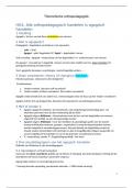
Skeletal muscle
- Most abundant (40%)
- Striated appearance- dark and light bands
- Voluntary muscle
- Multi-nucleated cells.
- Made up of muscle fibres.
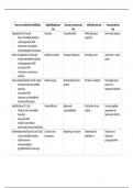
Muscle bundles of Fasciclea lot of Muscle fibres Myofibrilsthick and thin
filaments myosin and actin.
Group of muscle fibres is enveloped by perimysium which is a connective tissue.
Muscle cells are made up of long and cylindrical structures called myofibrils.
Myofibrils contains a Dark A band and a light I band.
1 thick filament is surrounded by 6 thin filaments.
Sarcomere is the functional unit of skeletal muscle- made up of A band and I band on either
side.
Functional unit is able to perform all the functions of the specific organ.
A band is made up of thick and thin filaments
I band is made up of ONLY thin filaments.
I Band A Band I Band
, Thick filament is made up of protein called myosin.
Thin filament structure
- Actin- spherical
The backbone of thin filament is made of actin molecules joined into two strands and
twisted together.
- Troponin- globular protein formed by 3 subunits.
One subunit interacts with calcium, one with actin and one reacts with tropomyosin.
- Tropomyosin- fibrous protein
Another protein present is Titin which allows the muscle to go to initial length after the
muscle is stretched.
Excitation- contraction coupling = the sequence of events that link the action
potential at the NMJ to the mechanical contraction of the muscle fibre.
ü What happens after the action potential travels to the skeletal muscle fibre?
The depolarisation at the surface of the muscle fibre travels down via transverse tubules to
the centre of the muscle fibres.
The surface of Transverse tubules contains specific receptors called dihydropyridine
receptors which are activated by the voltage.
This causes a permeability change within the sarcoplasmic reticulum which causes release of
Ca2+ to the cytosol.
- Most abundant (40%)
- Striated appearance- dark and light bands
- Voluntary muscle
- Multi-nucleated cells.
- Made up of muscle fibres.
Muscle bundles of Fasciclea lot of Muscle fibres Myofibrilsthick and thin
filaments myosin and actin.
Group of muscle fibres is enveloped by perimysium which is a connective tissue.
Muscle cells are made up of long and cylindrical structures called myofibrils.
Myofibrils contains a Dark A band and a light I band.
1 thick filament is surrounded by 6 thin filaments.
Sarcomere is the functional unit of skeletal muscle- made up of A band and I band on either
side.
Functional unit is able to perform all the functions of the specific organ.
A band is made up of thick and thin filaments
I band is made up of ONLY thin filaments.
I Band A Band I Band
, Thick filament is made up of protein called myosin.
Thin filament structure
- Actin- spherical
The backbone of thin filament is made of actin molecules joined into two strands and
twisted together.
- Troponin- globular protein formed by 3 subunits.
One subunit interacts with calcium, one with actin and one reacts with tropomyosin.
- Tropomyosin- fibrous protein
Another protein present is Titin which allows the muscle to go to initial length after the
muscle is stretched.
Excitation- contraction coupling = the sequence of events that link the action
potential at the NMJ to the mechanical contraction of the muscle fibre.
ü What happens after the action potential travels to the skeletal muscle fibre?
The depolarisation at the surface of the muscle fibre travels down via transverse tubules to
the centre of the muscle fibres.
The surface of Transverse tubules contains specific receptors called dihydropyridine
receptors which are activated by the voltage.
This causes a permeability change within the sarcoplasmic reticulum which causes release of
Ca2+ to the cytosol.