H1: Biomembrane structures
Biomembrane= phospholipid bilayer in which proteins are embedded
1) Plasma membrane defines cell + separates inside from
outside
2) Define intracellular organelles (nucleus, mitochondria,
lysosome)
3) Permeability barrier: prevent movement of water-soluble
substances from one side of membrane to other → different
composition inside compared to environment
4) Proteins embedded → different functions
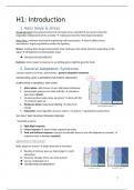
Fluid mosaic model of biomembranes
Phospholipid bilayer Phospholipids move laterally + spin
(~3nm thick) Non-covalent interaction between phospholipids + phospholipids-
proteins → important for strength of membrane
Hydrophobic core: lipids can’t move from one leaflet to another
Membrane proteins 1) Integral proteins: transmembrane
2) Lipid-anchored proteins: covalently attached hydrocarbon chain
3) Peripheral proteins: non-covalent interactions with integral
proteins/ membrane lipids
Contact with cytoskeleton
Prokaryotes vs eukaryotes
PROKARYOTES EUKARYOTES
1-2 µm 5-10 µm
Single PM PM: multitude of protein functions
NO internal membrane-limited Membrane-bound organelles → diverse
subcompartments proteins to carry out different functions
Proteins imbedded: ATP synthesis + PM proteins bind cytoskeleton
initiation DNA replication + membrane (=network of protein filaments that
transport proteins + receptors crisscrosses cytosol → provide
mechanical support for cellular
membranes + shape + cell movements
BEND + FLEX IN 3 DIMENSIONS WHILE MAINTAINING THEIR INTEGRITY → BECAUSE OF NON-
COVALENT INTERACTIONS BETWEEN LIPIDS AND PROTEINS
Fluid mosaic model of biomembranes= lipid bilayer behaves like two-dimensional fluid, with
individual lipid molecules able to move past one another + spin in place
Fluidity + flexibility allows:
1) Typical shape of organelles
2) Dynamic processes of membrane budding + fusion
1
, 1. Lipid bilayer: Composition and structural organization
Phospholipids= principal building blocks of biomembranes → most common: phosphoglycerides
➔ Amphipathic molecule: polar head group (strongly hydrophilic) + fatty-acid based
hydrocarbon tail (hydrophobic)
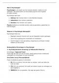
2. Phospholipids spontaneously form bilayers
Phospholipids in aqueous solution:
1) Spherical micelles: hydrophobic interior composed of fatty acyl
chains
2) Liposomes: phospholipid bilayer surrounding an aqueous center
3) Sheet-like phospholipid bilayer: 2 molecules thick, van der Waals
interactions inside of membrane (give membrane strength)
➔ Made by researchers: study membrane proteins in native
environment
Osmium tetroxide= stains phospholipid headgroups → appearance of railroad track
Type of structure formed depends on:
1) Length of fatty acyl chains
2) Degree of saturation
3) Temperature
➔ Fatty acyl chains aggregate + exclude water from core
MICELLES ARE RARELY FORMED FROM NATURAL PL!
1. Too bulky: not enough room in center of micelle to accommodate the chains
2. Use phospholipase → remove 1-2 of fatty acid chains (lysophospholipid)
3. Formation micelles
Leaflet Hydrophobic FA chains align tightly in center of bilayer → minimize
contact with water (3-4 nm thick hydrophobic core)
Interaction between HC chains: van der Waals interactions → stabilise
close packing
Interaction of polar head group with eachother + water: ionic + H-bonds
Phospholipid Basic structural unit of biological membranes:
bilayer Hydrophobic core: prevents water-soluble substances from crossing
Separates 2 aqueous solutions → permeability barrier
Defines cellular compartments + separates cells interior from world
➔ Longest cell membrane in history: 4.5 m (nerve cells of neck giraffe) + 40-50 m (tail nerves
Sauropods)
3. Phospholipid bilayers form sealed compartment surrounding
internal aqueous space
Important properties:
1) Impermeable to hydrophilic solutes
2) Stability: hydrophobic + van der Waals interactions between FA chains → maintain integrity
3) Spontaneous formation of sealed closed compartments → EDGE IS ENERGETICALLY
UNSTABLE
2
, Formation and study of pure phospholipid bilayers
1. Biological membrane with proteins imbedded
2. Treat with organic solvent (chloroform + methanol)
3. Solubilisation of PL + cholesterol → proteins + CH in insoluble
residue (centrifugation)
4. Method 1: Mechanically disperse lipids in water → spontaneously
form liposome
5. Method 2: Dissolve PL in solvent + apply to small hole in plastic
partition → formation planar bilayer → study physical properties of
bilayers
ALL MEMBRANES FORM CLOSED COMPARTMENTS!
Cytoplasmic face= oriented toward interior of cell (internal face)
Exoplasmic face= directed away from cytosol, in contact with external
environment (external face)
➔ Lumen is topologically equivalent to outside of cell!
Endocytosis= segment of PM buds inward toward the cytosol + pinches off a
separate vesicle → cytosolic face of PM remains facing cytosol + exoplasmic
face faces vesicle lumen
Exocytosis= intracellular vesicle fuses with plasma membrane → lumen
connects with extracellular medium
Endosymbiont hypothesis
• Nucleus + mitochondrion + chloroplast: 2 membranes separated by small intermembrane
space!
• Exoplasmic faces of inner/outer membranes border intermembrane space between them
Endosymbiont hypothesis= mitochondria + chloroplasts arose early in evolution of eukaryotic cells by
the engulfment of bacteria capable of oxidative phosphorylation/photosynthesis → 1 membrane
from host cell, 1 membrane from bacteria
➔ Origin of double membrane nucleus not known!
DIFFERENT CELL TYPES EXHIBIT VARIETY OF SHAPES, COMPLEMENTING A CELLS FUNCTION:
1) Smooth/flexible surface of erythrocyte → squeeze through narrow capillaries
2) Long, slender extension of PM (cilium/flagellum) → allows fluid to flow across surface of
sheet of cells + sperm cell to swim toward egg
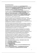
4. Biomembranes contain 3 principal classes of lipids
a) Phosphoglycerides
Phosphoglyceride= derivative of glycerol-3P + 2 esterified FA chains
(hydrophobic) + polar head group esterified to phosphate → amphipathic!
➔ FA vary in length + saturated/unsaturated
3
, - charged P-group & + charged OH groups on head group → strong interaction with water!
Neutral pH: PI + PS have net negative charge
Polar head groups pack together into bilayer structure
Phospholipases produce lysophospholipids: important signaling molecules!
Plasmalogens= group of phosphoglycerides with 1 FA chain attached to glycerol by ester linkeage + 1
attached by ether linkeage (greater chemical stability)→ in human brain + heart tissue
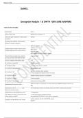
b) Sphingolipids
Sphingolipid= derived from sphingosine (amino alcohol with long
hydrocarbon chain) + long-chain FA attached via amide bond
1) Phosphate-based polar head group:
Sphingomyelin= phosphocholine attached to sphingosine
→ MAKES MIXED BILAYERS WITH PHOSPHOGLYCERIDES
2) Amphipathic glycolipids (polar head group=sugar):
Glucosylcerebroside= 1 glucose unit attached to sphingosine → 2-10% of total lipids in PM
(most abundant in nervous tissue)!
3) Sterols
Sterol= 4-ring isoprenoid-based hydrocarbon → OH-substituent on one ring: AMPHIPATHIC!
➔ No P-based head group: NOT PHOSPHOLIPIDS
➔ Too hydrophobic to form bilayer structure on their own!!
➔ Intercalate between PL molecules → influence membrane fluidity + provide
rigidity for mechanical support!
Cholesterol= precursor for several important bioactive molecule → bile acids (liver) + steroid
hormones (endocrine cells) + vitamin D (skin, kidneys)
➔ covalent addition to Hedgehog protein: signaling molecule in embryonic development
5. Lipids and proteins are laterally mobile in biomembranes
a) Rotate freely around their long axes
b) Diffuse laterally within each leaflet
BILAYER IS 100X MORE VISCOUS THAN WATER!
Phase transition= phospholipid membranes cooled <37°C change from fluid state (low viscosity) to
gel-like consistency
Gel-to-fluid transition
1. PL with long saturated FA chains → highly ordered gel-like bilayer
2. Heat disorders nonpolar tails
3. Transition from gel to fluid → tails overlap with eachother
4. Bilayer decreases in thickness
4
Biomembrane= phospholipid bilayer in which proteins are embedded
1) Plasma membrane defines cell + separates inside from
outside
2) Define intracellular organelles (nucleus, mitochondria,
lysosome)
3) Permeability barrier: prevent movement of water-soluble
substances from one side of membrane to other → different
composition inside compared to environment
4) Proteins embedded → different functions
Fluid mosaic model of biomembranes
Phospholipid bilayer Phospholipids move laterally + spin
(~3nm thick) Non-covalent interaction between phospholipids + phospholipids-
proteins → important for strength of membrane
Hydrophobic core: lipids can’t move from one leaflet to another
Membrane proteins 1) Integral proteins: transmembrane
2) Lipid-anchored proteins: covalently attached hydrocarbon chain
3) Peripheral proteins: non-covalent interactions with integral
proteins/ membrane lipids
Contact with cytoskeleton
Prokaryotes vs eukaryotes
PROKARYOTES EUKARYOTES
1-2 µm 5-10 µm
Single PM PM: multitude of protein functions
NO internal membrane-limited Membrane-bound organelles → diverse
subcompartments proteins to carry out different functions
Proteins imbedded: ATP synthesis + PM proteins bind cytoskeleton
initiation DNA replication + membrane (=network of protein filaments that
transport proteins + receptors crisscrosses cytosol → provide
mechanical support for cellular
membranes + shape + cell movements
BEND + FLEX IN 3 DIMENSIONS WHILE MAINTAINING THEIR INTEGRITY → BECAUSE OF NON-
COVALENT INTERACTIONS BETWEEN LIPIDS AND PROTEINS
Fluid mosaic model of biomembranes= lipid bilayer behaves like two-dimensional fluid, with
individual lipid molecules able to move past one another + spin in place
Fluidity + flexibility allows:
1) Typical shape of organelles
2) Dynamic processes of membrane budding + fusion
1
, 1. Lipid bilayer: Composition and structural organization
Phospholipids= principal building blocks of biomembranes → most common: phosphoglycerides
➔ Amphipathic molecule: polar head group (strongly hydrophilic) + fatty-acid based
hydrocarbon tail (hydrophobic)
2. Phospholipids spontaneously form bilayers
Phospholipids in aqueous solution:
1) Spherical micelles: hydrophobic interior composed of fatty acyl
chains
2) Liposomes: phospholipid bilayer surrounding an aqueous center
3) Sheet-like phospholipid bilayer: 2 molecules thick, van der Waals
interactions inside of membrane (give membrane strength)
➔ Made by researchers: study membrane proteins in native
environment
Osmium tetroxide= stains phospholipid headgroups → appearance of railroad track
Type of structure formed depends on:
1) Length of fatty acyl chains
2) Degree of saturation
3) Temperature
➔ Fatty acyl chains aggregate + exclude water from core
MICELLES ARE RARELY FORMED FROM NATURAL PL!
1. Too bulky: not enough room in center of micelle to accommodate the chains
2. Use phospholipase → remove 1-2 of fatty acid chains (lysophospholipid)
3. Formation micelles
Leaflet Hydrophobic FA chains align tightly in center of bilayer → minimize
contact with water (3-4 nm thick hydrophobic core)
Interaction between HC chains: van der Waals interactions → stabilise
close packing
Interaction of polar head group with eachother + water: ionic + H-bonds
Phospholipid Basic structural unit of biological membranes:
bilayer Hydrophobic core: prevents water-soluble substances from crossing
Separates 2 aqueous solutions → permeability barrier
Defines cellular compartments + separates cells interior from world
➔ Longest cell membrane in history: 4.5 m (nerve cells of neck giraffe) + 40-50 m (tail nerves
Sauropods)
3. Phospholipid bilayers form sealed compartment surrounding
internal aqueous space
Important properties:
1) Impermeable to hydrophilic solutes
2) Stability: hydrophobic + van der Waals interactions between FA chains → maintain integrity
3) Spontaneous formation of sealed closed compartments → EDGE IS ENERGETICALLY
UNSTABLE
2
, Formation and study of pure phospholipid bilayers
1. Biological membrane with proteins imbedded
2. Treat with organic solvent (chloroform + methanol)
3. Solubilisation of PL + cholesterol → proteins + CH in insoluble
residue (centrifugation)
4. Method 1: Mechanically disperse lipids in water → spontaneously
form liposome
5. Method 2: Dissolve PL in solvent + apply to small hole in plastic
partition → formation planar bilayer → study physical properties of
bilayers
ALL MEMBRANES FORM CLOSED COMPARTMENTS!
Cytoplasmic face= oriented toward interior of cell (internal face)
Exoplasmic face= directed away from cytosol, in contact with external
environment (external face)
➔ Lumen is topologically equivalent to outside of cell!
Endocytosis= segment of PM buds inward toward the cytosol + pinches off a
separate vesicle → cytosolic face of PM remains facing cytosol + exoplasmic
face faces vesicle lumen
Exocytosis= intracellular vesicle fuses with plasma membrane → lumen
connects with extracellular medium
Endosymbiont hypothesis
• Nucleus + mitochondrion + chloroplast: 2 membranes separated by small intermembrane
space!
• Exoplasmic faces of inner/outer membranes border intermembrane space between them
Endosymbiont hypothesis= mitochondria + chloroplasts arose early in evolution of eukaryotic cells by
the engulfment of bacteria capable of oxidative phosphorylation/photosynthesis → 1 membrane
from host cell, 1 membrane from bacteria
➔ Origin of double membrane nucleus not known!
DIFFERENT CELL TYPES EXHIBIT VARIETY OF SHAPES, COMPLEMENTING A CELLS FUNCTION:
1) Smooth/flexible surface of erythrocyte → squeeze through narrow capillaries
2) Long, slender extension of PM (cilium/flagellum) → allows fluid to flow across surface of
sheet of cells + sperm cell to swim toward egg
4. Biomembranes contain 3 principal classes of lipids
a) Phosphoglycerides
Phosphoglyceride= derivative of glycerol-3P + 2 esterified FA chains
(hydrophobic) + polar head group esterified to phosphate → amphipathic!
➔ FA vary in length + saturated/unsaturated
3
, - charged P-group & + charged OH groups on head group → strong interaction with water!
Neutral pH: PI + PS have net negative charge
Polar head groups pack together into bilayer structure
Phospholipases produce lysophospholipids: important signaling molecules!
Plasmalogens= group of phosphoglycerides with 1 FA chain attached to glycerol by ester linkeage + 1
attached by ether linkeage (greater chemical stability)→ in human brain + heart tissue
b) Sphingolipids
Sphingolipid= derived from sphingosine (amino alcohol with long
hydrocarbon chain) + long-chain FA attached via amide bond
1) Phosphate-based polar head group:
Sphingomyelin= phosphocholine attached to sphingosine
→ MAKES MIXED BILAYERS WITH PHOSPHOGLYCERIDES
2) Amphipathic glycolipids (polar head group=sugar):
Glucosylcerebroside= 1 glucose unit attached to sphingosine → 2-10% of total lipids in PM
(most abundant in nervous tissue)!
3) Sterols
Sterol= 4-ring isoprenoid-based hydrocarbon → OH-substituent on one ring: AMPHIPATHIC!
➔ No P-based head group: NOT PHOSPHOLIPIDS
➔ Too hydrophobic to form bilayer structure on their own!!
➔ Intercalate between PL molecules → influence membrane fluidity + provide
rigidity for mechanical support!
Cholesterol= precursor for several important bioactive molecule → bile acids (liver) + steroid
hormones (endocrine cells) + vitamin D (skin, kidneys)
➔ covalent addition to Hedgehog protein: signaling molecule in embryonic development
5. Lipids and proteins are laterally mobile in biomembranes
a) Rotate freely around their long axes
b) Diffuse laterally within each leaflet
BILAYER IS 100X MORE VISCOUS THAN WATER!
Phase transition= phospholipid membranes cooled <37°C change from fluid state (low viscosity) to
gel-like consistency
Gel-to-fluid transition
1. PL with long saturated FA chains → highly ordered gel-like bilayer
2. Heat disorders nonpolar tails
3. Transition from gel to fluid → tails overlap with eachother
4. Bilayer decreases in thickness
4