Inhoudsopgave
PART I: PHYSICAL PRINCIPLES MR IMAGING .................................................................................... 3
WHAT IS MRI? ................................................................................................................................... 3
IMAGES:..................................................................................................................................................... 3
SYNOPSYS .................................................................................................................................................. 4
KIND OF NUCLEI........................................................................................................................................... 4
MOTION OF Μ IN MAGNETIC FIELD B0 ............................................................................................................. 5
POTENTIAL INTERACTION ENERGIES: ................................................................................................................ 6
NETTO MAGNETISATION ............................................................................................................................... 7
MOTION OF M IN B0 FIELD ............................................................................................................................ 7
ROTATING FRAME OF REFERENCE.................................................................................................................... 8
HOW TO PERTURB M? (PERTURBATION = VERSTORING) ..................................................................................... 8
EXPERIMENTAL SET-UP MICE AND STARLING ..................................................................................................... 9
RF SIGNAL.......................................................................................................................................... 9
AFTER THE RF PULSE .................................................................................................................................... 9
DETECTION OF THE SIGNAL .......................................................................................................................... 10
RELAXATION ..................................................................................................................................... 11
T1 RELAXATION ......................................................................................................................................... 11
T2 RELAXATION ......................................................................................................................................... 13
T2 AND T2* RELAXATION............................................................................................................................ 14
SEQUENCES ...................................................................................................................................... 15
GRADIENT-ECHO SEQUENCE ........................................................................................................................ 15
SPIN-ECHO SEQUENCE: T2 RELAXATION .......................................................................................................... 16
CARR PURCELL MEIBOOM GILL SPIN ECHO SEQUENCE ...................................................................................... 17
SPIN-ECHO IMAGE CONTRAST ............................................................................................................... 18
T2 CONTRAST ........................................................................................................................................... 19
T1 CONTRAST ........................................................................................................................................... 19
PROTON DENSITY WEIGHTED ....................................................................................................................... 19
INVERSION RECOVERY SEQUENCE: T1-WEIGHING............................................................................................. 19
PART I: MR IMAGE RECONSTRUCTION ........................................................................................... 20
FROM RAW DATA – IN K-SPACE ............................................................................................................. 20
FOURIER TRANSFORMATION................................................................................................................. 21
INTRODUCTION ......................................................................................................................................... 21
FT DEFENITIONS ........................................................................................................................................ 22
VERTICAL PROJECTION ................................................................................................................................ 22
THE + AND – FREQUENCY PROBLEM .............................................................................................................. 23
FOURIER PAIR............................................................................................................................................ 23
CONVOLUTION THEOREME .......................................................................................................................... 23
DIGITAL FT ............................................................................................................................................... 23
2D FT ..................................................................................................................................................... 23
AN INTRODUCTION IN CREATING AN IMAGE ............................................................................................. 24
THE MR-SEQUENCE ................................................................................................................................... 24
1
,EXCITATION .............................................................................................................................................. 24
ACQUISITION ............................................................................................................................................ 24
RF-PULSE ................................................................................................................................................. 24
MAGNETIC FIELD GRADIENT ........................................................................................................................ 25
MR-SYSTEM ............................................................................................................................................. 26
PHASE ENCODING GRADIENT GY ................................................................................................................... 32
MRIMAGING OPTIONS........................................................................................................................ 33
MRI : 2D-FOURIER IMAGING ...................................................................................................................... 33
MRI : SINGLE-SLICE IMAGING ...................................................................................................................... 35
MRI : MULTI-SLICE IMAGING ....................................................................................................................... 35
MRI : 3D-IMAGING ................................................................................................................................... 35
SIGNAL AND NOISE .................................................................................................................................... 35
IMAGE CHARACTERISTICS ..................................................................................................................... 36
FIELD OF VIEW (FOV) ................................................................................................................................. 36
IMAGE MATRIX .......................................................................................................................................... 36
SPATIAL RESOLUTION .................................................................................................................................. 36
TEMPORAL RESOLUTION .............................................................................................................................. 36
SIGNAL-TO-NOISE RATIO ............................................................................................................................. 36
CONTRAST TO NOISE RATIO ......................................................................................................................... 36
CONTRAST AND BRIGHTNESS........................................................................................................................ 37
PART II: ADVANCED MRI................................................................................................................ 38
FLOW EFFECTS, FLOW IMAGING, ANGIOGRAPHY ....................................................................................... 38
FLOW EFFECTS ON MR IMAGES .................................................................................................................... 38
TOF EFFECTS: FLOW OUT OF PLANE .............................................................................................................. 38
TOF EFFECTS: FLOW OUT OF PLANE: GE (TR) ................................................................................................. 39
TOF EFFECTS: FLOW OUT OF PLANE: SE ......................................................................................................... 40
TOF EFFECTS: FLOW WITHIN THE PLANE ........................................................................................................ 40
EFFECT MOTION – DURING IMAGING ............................................................................................................. 42
PHASE EFFECTS .......................................................................................................................................... 42
TIME-OF-FLIGHT MR ANGIOGRAPHY ............................................................................................................. 43
TOF MR ANGIOGRAPHY: STATIC MATERIAL SUPPRESSION ................................................................................. 43
TOF MR ANGIOGRAPHY: FLOW SENSITISATION ............................................................................................... 43
CONTRAST ENHANCED ANGIOGRAPHY ........................................................................................................... 44
PRINCIPLES OF 3D GD-ENHANCED MR ANGIOGRAPHY ..................................................................................... 45
QUANTITATIVE FLOW MR IMAGING: INTRODUCTION ....................................................................................... 48
PHASE CONTRAST IMAGES ........................................................................................................................... 48
2D PCA IMAGING...................................................................................................................................... 51
3D PCA IMAGING...................................................................................................................................... 51
PHASE CONTRAST ANGIOGRAPHY + VELOCIMETRY ........................................................................................... 51
DIFFUSION ....................................................................................................................................... 51
PHYSICAL PRINCIPLES .................................................................................................................................. 51
RESTRICTED AND FREE DIFFUSION ................................................................................................................. 53
DIFFUSION AND DIFFUSION WEIGHTING IN MRI .............................................................................................. 53
ADC MAPS ............................................................................................................................................... 55
PARAMETERS INFLUENCING DIFFUSION MEASUREMENTS .................................................................................. 56
BIOMEDICAL APPLICATION ........................................................................................................................... 57
CONTRAST AGENTS & MEMRI ............................................................................................................ 61
PERFUSION – BOLUS TRACKING ............................................................................................................. 62
PERFUSION – ASL PET FMRI ............................................................................................................... 62
2
,Preclinical and clinical imaging with focus
on neurology
Part I: Physical Principles MR imaging
We use waves, and these waves will interact with your body. What you see/detect is the interaction. We will
explain in debt al different interactions. ct also uses waves EM to look at nuclei. another part clinically imprtant
is ultrasound. We dont use it to look at human brain because of the skull. Its very hard to get through the skull.
there is too much reflection. only with babies it is possible because they have a soft fontanel
EM spectrum:
C=f*λ
Cvacuum = 3 * 108 m/s
Energy of a photon: E = h * f
H = Planck’s constant (6.63*10-34 Js)
All the types of waves. an EM wave is like a foton and
they each have an energy and the energy is given by
the wave. So if you have low frequency there is a low energystate. Mri is in the top row so has low frequency and
such a low energy as compared to EM-waves used for RX and CT.
What is the difference? What do we have to consider ? The health of the patient, radioprotection. Only the part
we want to see, needs to be radiated with waves, also the nurses and doctors need to be protected. This is not
a problem with mri. Because the ionization is the real danger, which isn't used in MRI.
What is MRI?
- Proton density hydrogen nuclei density (number of protons) : source of analysing: more protons =
higher signal.
- Also interaction of these protons with surroundings (T1, T2, ..)
- We don’t have to kill the animal to gain information
- Different imaging sequences provide
o Anatomical information
o physiological information
o functional information
o Molecular information, migration of labeled stem cells
o few other nuclei may be used for specialised imaging purposes (e.g. 13C, 31P).
- MRI is particularly suited to imaging differences between soft tissues, such as in the head, neck and
spinal regions of the body. à not really used for bone
Aperture is small so you can only put small animals in it, on which we study neurological topics. We see horizontal
images of the head of the mouse and with MRI you can get different images with different contrast. With MRI
we need to specify what you show.
What is different with the CT? At the CT the bones are always bright. It's the density that we see on the CT image.
In MRI it's always different what you get to see, different contrasts and features. This are anatomical images. You
can measure volumes.
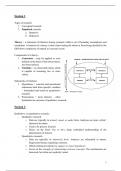
Images:
The images have a slice thickness à divide à small volume element
2 D à pixel : intensity, grey level: = a picture element. It's 2-Dimensional.
An MR image is compromised of pixels. And we derive the pixel from a
voxel.
3
, 3 D à Voxel : property parameter in the volume: a volume element. It's 3-Dimensional. like the human brain
for example. An image (pixels) is a slice through the 3D-object.
Here you see the patient, its a 3D object. With mri you make an image, existing oft pixels. the information in
your pixel, comes from a splice. You select a splice in the brain of the patient or animal. Each of those volumes,
will contribute to a watermolecule, and hydrogen nuclei. From the hydrogen nuclei or interactions you get
from your signal. Dpending on the contrast you want, you get a very high or low intensity(density?)
Our image is 2D. Pixel is a picture element which comes from a voxel which is a volume element, has a grey
level and the instensity is related to what kind of image you want to show, which can't of contrast
The pixel represents the signal from a voxel. 1 vlakte van de kubus
Synopsys
what do you need for MRI? you need a very big magnetic field. How do you create a magnetic field? Electrical
current, going through the coil. Right hand rule (duim = richting elektrisch veld en hand = richting stroom),
follow the direction of the current, the thumb will show you the direction of the magnetic field. 3 tesla for
humans. For small animals we use 11.5 T. This can be an elektricalmotive coil "you plug in and you plug out".
Today we use superconducting magnet, not the previous one.
Why? Because they don't have a resistance. If you cool down, the resistance of the conductor reduces. Up to 4
kelvin there is no elektrical resistance. Everything is submerged. You can remove it and the current that was
there keeps on running all the time, also in the evening. That's different part in the mri. Not the waves but the
attractive forces that are created by the magnetic force. We change the equilibrium by transmitting
radiowaves in the subject. The subject gets in the scanner, and around the part of the body where we want tot
take the image, has a certain coil, waves for short time will be emitting. And outside of the body itself, the
electrowaves are transmitted, which are coming out of the body. this is the source of our image. De data we
convert to an image.
Exposure of subject to big static magnetic field
0.2 (vet) à 0.5T à 3T (human) à 11.5 T (small animal)
(superconducting magnet – coil immersed in liquid He at 4K so very low temperature à
the resistance will also drop. The helium will be consumed a little bit so you will need to
refill it. Some older scanners have also Nitro in them à accidents can happen
! the scanner is always on!
- Dedicated coil (part body): Transmit radio waves into subject [2~10 ms] à very breef moment
- Turn off radio wave transmitter
- Apply time-varying magnetic fields (position encoding): where does it come from
- Receive radio waves re-transmitted by subject with the same coil
- Data will be very complex!
- Convert measured RF data to image
Kind of nuclei
nMRI: nuclear imaging: had a bad connotation so now it is just MRI
Now we will see the basic of the mri. A proton you can use to make an mri. there are different characteristics a
nucleus needs to have. All the nuclei who have an altered number of proton and neutron have a spin. You see a
list of elements that can be used for mri. The hydrogen atom has the highest natural abundace in the body. You
can also do metrosprectomy. Fluor is used as a contrast agent, you measure the density of your fluor nuclei.
sadly we can't measure oxygen directly. The abundance is very important, If it is low there will not be enough
signal/ it will not exceed the noice signal.
H+ has 1 proton. You can also use other nuclei. E.G.: phosphor for metabolism
We can not look at Oxygen or C12 à Because they have an even number of protons or neutrons !!
Nucleus needs to have 2 properties:
- Spin
- charge
Nuclei are made of protons and neutrons:
- Both have spin 1⁄2
- Protons have charge
à This means every atom will have a magnetic moment: µ
Add those all together and you will get a magnetic field.
Pairs of spins tend to cancel, so only atoms with an odd number of protons or neutrons have spin
4
PART I: PHYSICAL PRINCIPLES MR IMAGING .................................................................................... 3
WHAT IS MRI? ................................................................................................................................... 3
IMAGES:..................................................................................................................................................... 3
SYNOPSYS .................................................................................................................................................. 4
KIND OF NUCLEI........................................................................................................................................... 4
MOTION OF Μ IN MAGNETIC FIELD B0 ............................................................................................................. 5
POTENTIAL INTERACTION ENERGIES: ................................................................................................................ 6
NETTO MAGNETISATION ............................................................................................................................... 7
MOTION OF M IN B0 FIELD ............................................................................................................................ 7
ROTATING FRAME OF REFERENCE.................................................................................................................... 8
HOW TO PERTURB M? (PERTURBATION = VERSTORING) ..................................................................................... 8
EXPERIMENTAL SET-UP MICE AND STARLING ..................................................................................................... 9
RF SIGNAL.......................................................................................................................................... 9
AFTER THE RF PULSE .................................................................................................................................... 9
DETECTION OF THE SIGNAL .......................................................................................................................... 10
RELAXATION ..................................................................................................................................... 11
T1 RELAXATION ......................................................................................................................................... 11
T2 RELAXATION ......................................................................................................................................... 13
T2 AND T2* RELAXATION............................................................................................................................ 14
SEQUENCES ...................................................................................................................................... 15
GRADIENT-ECHO SEQUENCE ........................................................................................................................ 15
SPIN-ECHO SEQUENCE: T2 RELAXATION .......................................................................................................... 16
CARR PURCELL MEIBOOM GILL SPIN ECHO SEQUENCE ...................................................................................... 17
SPIN-ECHO IMAGE CONTRAST ............................................................................................................... 18
T2 CONTRAST ........................................................................................................................................... 19
T1 CONTRAST ........................................................................................................................................... 19
PROTON DENSITY WEIGHTED ....................................................................................................................... 19
INVERSION RECOVERY SEQUENCE: T1-WEIGHING............................................................................................. 19
PART I: MR IMAGE RECONSTRUCTION ........................................................................................... 20
FROM RAW DATA – IN K-SPACE ............................................................................................................. 20
FOURIER TRANSFORMATION................................................................................................................. 21
INTRODUCTION ......................................................................................................................................... 21
FT DEFENITIONS ........................................................................................................................................ 22
VERTICAL PROJECTION ................................................................................................................................ 22
THE + AND – FREQUENCY PROBLEM .............................................................................................................. 23
FOURIER PAIR............................................................................................................................................ 23
CONVOLUTION THEOREME .......................................................................................................................... 23
DIGITAL FT ............................................................................................................................................... 23
2D FT ..................................................................................................................................................... 23
AN INTRODUCTION IN CREATING AN IMAGE ............................................................................................. 24
THE MR-SEQUENCE ................................................................................................................................... 24
1
,EXCITATION .............................................................................................................................................. 24
ACQUISITION ............................................................................................................................................ 24
RF-PULSE ................................................................................................................................................. 24
MAGNETIC FIELD GRADIENT ........................................................................................................................ 25
MR-SYSTEM ............................................................................................................................................. 26
PHASE ENCODING GRADIENT GY ................................................................................................................... 32
MRIMAGING OPTIONS........................................................................................................................ 33
MRI : 2D-FOURIER IMAGING ...................................................................................................................... 33
MRI : SINGLE-SLICE IMAGING ...................................................................................................................... 35
MRI : MULTI-SLICE IMAGING ....................................................................................................................... 35
MRI : 3D-IMAGING ................................................................................................................................... 35
SIGNAL AND NOISE .................................................................................................................................... 35
IMAGE CHARACTERISTICS ..................................................................................................................... 36
FIELD OF VIEW (FOV) ................................................................................................................................. 36
IMAGE MATRIX .......................................................................................................................................... 36
SPATIAL RESOLUTION .................................................................................................................................. 36
TEMPORAL RESOLUTION .............................................................................................................................. 36
SIGNAL-TO-NOISE RATIO ............................................................................................................................. 36
CONTRAST TO NOISE RATIO ......................................................................................................................... 36
CONTRAST AND BRIGHTNESS........................................................................................................................ 37
PART II: ADVANCED MRI................................................................................................................ 38
FLOW EFFECTS, FLOW IMAGING, ANGIOGRAPHY ....................................................................................... 38
FLOW EFFECTS ON MR IMAGES .................................................................................................................... 38
TOF EFFECTS: FLOW OUT OF PLANE .............................................................................................................. 38
TOF EFFECTS: FLOW OUT OF PLANE: GE (TR) ................................................................................................. 39
TOF EFFECTS: FLOW OUT OF PLANE: SE ......................................................................................................... 40
TOF EFFECTS: FLOW WITHIN THE PLANE ........................................................................................................ 40
EFFECT MOTION – DURING IMAGING ............................................................................................................. 42
PHASE EFFECTS .......................................................................................................................................... 42
TIME-OF-FLIGHT MR ANGIOGRAPHY ............................................................................................................. 43
TOF MR ANGIOGRAPHY: STATIC MATERIAL SUPPRESSION ................................................................................. 43
TOF MR ANGIOGRAPHY: FLOW SENSITISATION ............................................................................................... 43
CONTRAST ENHANCED ANGIOGRAPHY ........................................................................................................... 44
PRINCIPLES OF 3D GD-ENHANCED MR ANGIOGRAPHY ..................................................................................... 45
QUANTITATIVE FLOW MR IMAGING: INTRODUCTION ....................................................................................... 48
PHASE CONTRAST IMAGES ........................................................................................................................... 48
2D PCA IMAGING...................................................................................................................................... 51
3D PCA IMAGING...................................................................................................................................... 51
PHASE CONTRAST ANGIOGRAPHY + VELOCIMETRY ........................................................................................... 51
DIFFUSION ....................................................................................................................................... 51
PHYSICAL PRINCIPLES .................................................................................................................................. 51
RESTRICTED AND FREE DIFFUSION ................................................................................................................. 53
DIFFUSION AND DIFFUSION WEIGHTING IN MRI .............................................................................................. 53
ADC MAPS ............................................................................................................................................... 55
PARAMETERS INFLUENCING DIFFUSION MEASUREMENTS .................................................................................. 56
BIOMEDICAL APPLICATION ........................................................................................................................... 57
CONTRAST AGENTS & MEMRI ............................................................................................................ 61
PERFUSION – BOLUS TRACKING ............................................................................................................. 62
PERFUSION – ASL PET FMRI ............................................................................................................... 62
2
,Preclinical and clinical imaging with focus
on neurology
Part I: Physical Principles MR imaging
We use waves, and these waves will interact with your body. What you see/detect is the interaction. We will
explain in debt al different interactions. ct also uses waves EM to look at nuclei. another part clinically imprtant
is ultrasound. We dont use it to look at human brain because of the skull. Its very hard to get through the skull.
there is too much reflection. only with babies it is possible because they have a soft fontanel
EM spectrum:
C=f*λ
Cvacuum = 3 * 108 m/s
Energy of a photon: E = h * f
H = Planck’s constant (6.63*10-34 Js)
All the types of waves. an EM wave is like a foton and
they each have an energy and the energy is given by
the wave. So if you have low frequency there is a low energystate. Mri is in the top row so has low frequency and
such a low energy as compared to EM-waves used for RX and CT.
What is the difference? What do we have to consider ? The health of the patient, radioprotection. Only the part
we want to see, needs to be radiated with waves, also the nurses and doctors need to be protected. This is not
a problem with mri. Because the ionization is the real danger, which isn't used in MRI.
What is MRI?
- Proton density hydrogen nuclei density (number of protons) : source of analysing: more protons =
higher signal.
- Also interaction of these protons with surroundings (T1, T2, ..)
- We don’t have to kill the animal to gain information
- Different imaging sequences provide
o Anatomical information
o physiological information
o functional information
o Molecular information, migration of labeled stem cells
o few other nuclei may be used for specialised imaging purposes (e.g. 13C, 31P).
- MRI is particularly suited to imaging differences between soft tissues, such as in the head, neck and
spinal regions of the body. à not really used for bone
Aperture is small so you can only put small animals in it, on which we study neurological topics. We see horizontal
images of the head of the mouse and with MRI you can get different images with different contrast. With MRI
we need to specify what you show.
What is different with the CT? At the CT the bones are always bright. It's the density that we see on the CT image.
In MRI it's always different what you get to see, different contrasts and features. This are anatomical images. You
can measure volumes.
Images:
The images have a slice thickness à divide à small volume element
2 D à pixel : intensity, grey level: = a picture element. It's 2-Dimensional.
An MR image is compromised of pixels. And we derive the pixel from a
voxel.
3
, 3 D à Voxel : property parameter in the volume: a volume element. It's 3-Dimensional. like the human brain
for example. An image (pixels) is a slice through the 3D-object.
Here you see the patient, its a 3D object. With mri you make an image, existing oft pixels. the information in
your pixel, comes from a splice. You select a splice in the brain of the patient or animal. Each of those volumes,
will contribute to a watermolecule, and hydrogen nuclei. From the hydrogen nuclei or interactions you get
from your signal. Dpending on the contrast you want, you get a very high or low intensity(density?)
Our image is 2D. Pixel is a picture element which comes from a voxel which is a volume element, has a grey
level and the instensity is related to what kind of image you want to show, which can't of contrast
The pixel represents the signal from a voxel. 1 vlakte van de kubus
Synopsys
what do you need for MRI? you need a very big magnetic field. How do you create a magnetic field? Electrical
current, going through the coil. Right hand rule (duim = richting elektrisch veld en hand = richting stroom),
follow the direction of the current, the thumb will show you the direction of the magnetic field. 3 tesla for
humans. For small animals we use 11.5 T. This can be an elektricalmotive coil "you plug in and you plug out".
Today we use superconducting magnet, not the previous one.
Why? Because they don't have a resistance. If you cool down, the resistance of the conductor reduces. Up to 4
kelvin there is no elektrical resistance. Everything is submerged. You can remove it and the current that was
there keeps on running all the time, also in the evening. That's different part in the mri. Not the waves but the
attractive forces that are created by the magnetic force. We change the equilibrium by transmitting
radiowaves in the subject. The subject gets in the scanner, and around the part of the body where we want tot
take the image, has a certain coil, waves for short time will be emitting. And outside of the body itself, the
electrowaves are transmitted, which are coming out of the body. this is the source of our image. De data we
convert to an image.
Exposure of subject to big static magnetic field
0.2 (vet) à 0.5T à 3T (human) à 11.5 T (small animal)
(superconducting magnet – coil immersed in liquid He at 4K so very low temperature à
the resistance will also drop. The helium will be consumed a little bit so you will need to
refill it. Some older scanners have also Nitro in them à accidents can happen
! the scanner is always on!
- Dedicated coil (part body): Transmit radio waves into subject [2~10 ms] à very breef moment
- Turn off radio wave transmitter
- Apply time-varying magnetic fields (position encoding): where does it come from
- Receive radio waves re-transmitted by subject with the same coil
- Data will be very complex!
- Convert measured RF data to image
Kind of nuclei
nMRI: nuclear imaging: had a bad connotation so now it is just MRI
Now we will see the basic of the mri. A proton you can use to make an mri. there are different characteristics a
nucleus needs to have. All the nuclei who have an altered number of proton and neutron have a spin. You see a
list of elements that can be used for mri. The hydrogen atom has the highest natural abundace in the body. You
can also do metrosprectomy. Fluor is used as a contrast agent, you measure the density of your fluor nuclei.
sadly we can't measure oxygen directly. The abundance is very important, If it is low there will not be enough
signal/ it will not exceed the noice signal.
H+ has 1 proton. You can also use other nuclei. E.G.: phosphor for metabolism
We can not look at Oxygen or C12 à Because they have an even number of protons or neutrons !!
Nucleus needs to have 2 properties:
- Spin
- charge
Nuclei are made of protons and neutrons:
- Both have spin 1⁄2
- Protons have charge
à This means every atom will have a magnetic moment: µ
Add those all together and you will get a magnetic field.
Pairs of spins tend to cancel, so only atoms with an odd number of protons or neutrons have spin
4