1-Conventional X-ray imaging
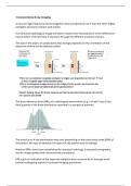
X-rays are high-frequency electromagnetic waves produced in an X-ray tube when highly
energetic electrons interact with matter.
Conventional radiological image formation results from the projection of the differential
transmission of the primary X-ray beam through the different anatomic tissues.
The size of the object on projections also strongly depends on the orientation of the
objective relative tot the detector plane!
The dose reference level (DRL) of a radiological examination (e.g. a chest X-ray) is the
third quartile of the dose distribution reported in a sample of patients.
The dose for an X-ray examination may vary depending on the body mass index (BMI) of
the patient, the type of detector, the type of X-ray system and its settings.
National DRLs have been established for standard radiology, computed tomography,
and for image-guided and interventional procedures.
DRLs give an indication of the expected radiation dose received by an average-sized
patient undergoing a given X-ray based imaging procedure.
,DRLs are a tool to optimize medical imaging procedures using ionizing radiation
DRL are not dose limits!
2-Ultrasound
Sonography is a non-invasive painless procedure, which uses ultrasound waves to
produce images of organs, blood vessels or soft tissues for medical analysis. The terms
sonography and ultrasound are often used interchangeably. A sonogram is an image
generated by ultrasound.
Ultrasound waves have frequencies higher than the upper limit of human hearing. In
medical ultrasound, frequencies typically fall in the 1 to 20 MHz range, while the upper
limit of human hearing is around 20 kHz.
The basic ultrasound principle:
- An ultrasound transducer emits an ultrasound signal.
- The transducer listens for the echo generated by the structures that the wave
encounters.
- The echo is turned into an image based on characteristics of the echo, such as
timing, amplitude and frequency.
Ultrasound interacts with tissues in different ways:
- Reflection - waves are reflected back to the transducer
- Absorption – waves are absorbed by the tissue and the energy is converted to
heat
- Scattering – waves are reflected in multiple different directions
- Refraction – the direction of waves is changed
Each type of tissue has a particular impedance – a resistance to the propagation of
sound which depends on the tissue density and the speed of sound in the tissue.
The amount of generated reflection depends on differences in impedance between
tissues.
Structures that elicit a powerful echo appear bright on our screen – we call them
hyperechoic
Structures that elicit a weak echo appear dark on our screen – we call them hypoechoic
Structures that elicit an echo similar to their surrounding structure are called isoechoic
,Strengths and limitations of ultrasound vary greatly with different applications.
Below is an outline of general strengths and limitations of ultrasound as opposed to
other imaging modalities, such as CT and MRI, that one needs to consider when
choosing between modalities.
Strengths: Limitations:
- Low cost - Highly operator dependant
- High availability - Highly patient dependant
- High portability - Difficult to reproduce
- Safe and non-invasive - Poor penetration in air and bone
- Fast
- Dynamic
3-Computed tomography
CT scanners include the following main components:
- An X-ray tube
- A gantry with detectors which are sensitive to X rays. The X ray detectors are
located directly opposite the X ray source.
- A computer
Images are generated using the same principle as in conventional radiography (see
chapter on conventional radiography), however, the X ray source rotates around the
gantry (unlike conventional X ray radiography, which uses a fixed X ray tube)
During a CT examination, the patient lies on a bed that moves through the gantry while
the X ray tube rotates around the patient.
, Depending on the indication, oral or intravenous or contrast media can be administered.
X-rays are high-frequency electromagnetic waves produced in an X-ray tube when highly
energetic electrons interact with matter.
Conventional radiological image formation results from the projection of the differential
transmission of the primary X-ray beam through the different anatomic tissues.
The size of the object on projections also strongly depends on the orientation of the
objective relative tot the detector plane!
The dose reference level (DRL) of a radiological examination (e.g. a chest X-ray) is the
third quartile of the dose distribution reported in a sample of patients.
The dose for an X-ray examination may vary depending on the body mass index (BMI) of
the patient, the type of detector, the type of X-ray system and its settings.
National DRLs have been established for standard radiology, computed tomography,
and for image-guided and interventional procedures.
DRLs give an indication of the expected radiation dose received by an average-sized
patient undergoing a given X-ray based imaging procedure.
,DRLs are a tool to optimize medical imaging procedures using ionizing radiation
DRL are not dose limits!
2-Ultrasound
Sonography is a non-invasive painless procedure, which uses ultrasound waves to
produce images of organs, blood vessels or soft tissues for medical analysis. The terms
sonography and ultrasound are often used interchangeably. A sonogram is an image
generated by ultrasound.
Ultrasound waves have frequencies higher than the upper limit of human hearing. In
medical ultrasound, frequencies typically fall in the 1 to 20 MHz range, while the upper
limit of human hearing is around 20 kHz.
The basic ultrasound principle:
- An ultrasound transducer emits an ultrasound signal.
- The transducer listens for the echo generated by the structures that the wave
encounters.
- The echo is turned into an image based on characteristics of the echo, such as
timing, amplitude and frequency.
Ultrasound interacts with tissues in different ways:
- Reflection - waves are reflected back to the transducer
- Absorption – waves are absorbed by the tissue and the energy is converted to
heat
- Scattering – waves are reflected in multiple different directions
- Refraction – the direction of waves is changed
Each type of tissue has a particular impedance – a resistance to the propagation of
sound which depends on the tissue density and the speed of sound in the tissue.
The amount of generated reflection depends on differences in impedance between
tissues.
Structures that elicit a powerful echo appear bright on our screen – we call them
hyperechoic
Structures that elicit a weak echo appear dark on our screen – we call them hypoechoic
Structures that elicit an echo similar to their surrounding structure are called isoechoic
,Strengths and limitations of ultrasound vary greatly with different applications.
Below is an outline of general strengths and limitations of ultrasound as opposed to
other imaging modalities, such as CT and MRI, that one needs to consider when
choosing between modalities.
Strengths: Limitations:
- Low cost - Highly operator dependant
- High availability - Highly patient dependant
- High portability - Difficult to reproduce
- Safe and non-invasive - Poor penetration in air and bone
- Fast
- Dynamic
3-Computed tomography
CT scanners include the following main components:
- An X-ray tube
- A gantry with detectors which are sensitive to X rays. The X ray detectors are
located directly opposite the X ray source.
- A computer
Images are generated using the same principle as in conventional radiography (see
chapter on conventional radiography), however, the X ray source rotates around the
gantry (unlike conventional X ray radiography, which uses a fixed X ray tube)
During a CT examination, the patient lies on a bed that moves through the gantry while
the X ray tube rotates around the patient.
, Depending on the indication, oral or intravenous or contrast media can be administered.