MS 20454151
How cell division in eukaryotic cells contributes to genetic variation
Onion root tip experiment to observe mitosis under a microscope
Mitosis and meiosis are both types of cell division. We can observe
mitosis or meiosis with a microscope by placing small pieces of
onion/garlic root tips under a microscope and analysing it. The main
theory for this experiment is simply that cell division is a necessary step in
anything cell related.
I will be going over the onion root tips experiment.
There are many four main stages in mitosis and those are, prophase,
metaphase, anaphase and telophase. The tip of the onion roots contains
meristematic cells which means they have no specific function making it
a good raw material for scientific studies. On top of that onion is a
monocotyledonous plant and they are known to hold large chromosomes.
Equipment list
- Light microscope
- Onion root peel
- Acetocarmine stain
- Water
- Bunsen burner
- N/10 hydrochloric acid
- Filter paper
- Coverslip
- Aceto alcohol (combination of acetic acid and ethanol in a 1:3 ratio)
- Watch glass
- Needle
- Vial
- Dropper
- Forceps
- Blade
Method
1. Get your onion and using your blade cut off the dry roots of the
onion. Place the root into a beaker filled with water so that the root
tips can grow. This may take a few days.
2. Once the root has grown in length, cut off around 3cm of the newly
grown roots and place them in your watch glass.
3. Using forceps, you should now transfer it to a vial containing an the
premade aceto alcohol.
4. Once again let the root tips in the vial for at least a day.
P a g e 1 | 10
, MS 20454151
5. Once you’ve waited long enough, use the forceps to pick a root and
put it on a glass slide ensuring you’re not contaminating the glass
slide with anything such as leaving fingerprints where the subject is
6. . Drop some N10/ hcl onto the onion root tip, as well as 2 to 3 drops
of the acetocarmine stain.
7. Turn on the Bunsen burner and lightly but not too much so that the
stain dries.
8. If you accidentally put too much stain onto the glass, you can use
filter paper to remove some of it.
9. Place the part of the root tip that doesn’t have too much stain on it
so it can be visualised clearer.
10. Add a small droplet of water onto the slide and then put a
cover slip over top of it. Very gently use the blunt side of a needle to
press down the cover slip.
11. You’re now left with an onion root tip cell slide that is ready to
be examined.
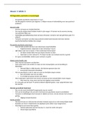
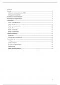
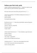
This is what the onion root looks like at 400x magnification.
P a g e 2 | 10
How cell division in eukaryotic cells contributes to genetic variation
Onion root tip experiment to observe mitosis under a microscope
Mitosis and meiosis are both types of cell division. We can observe
mitosis or meiosis with a microscope by placing small pieces of
onion/garlic root tips under a microscope and analysing it. The main
theory for this experiment is simply that cell division is a necessary step in
anything cell related.
I will be going over the onion root tips experiment.
There are many four main stages in mitosis and those are, prophase,
metaphase, anaphase and telophase. The tip of the onion roots contains
meristematic cells which means they have no specific function making it
a good raw material for scientific studies. On top of that onion is a
monocotyledonous plant and they are known to hold large chromosomes.
Equipment list
- Light microscope
- Onion root peel
- Acetocarmine stain
- Water
- Bunsen burner
- N/10 hydrochloric acid
- Filter paper
- Coverslip
- Aceto alcohol (combination of acetic acid and ethanol in a 1:3 ratio)
- Watch glass
- Needle
- Vial
- Dropper
- Forceps
- Blade
Method
1. Get your onion and using your blade cut off the dry roots of the
onion. Place the root into a beaker filled with water so that the root
tips can grow. This may take a few days.
2. Once the root has grown in length, cut off around 3cm of the newly
grown roots and place them in your watch glass.
3. Using forceps, you should now transfer it to a vial containing an the
premade aceto alcohol.
4. Once again let the root tips in the vial for at least a day.
P a g e 1 | 10
, MS 20454151
5. Once you’ve waited long enough, use the forceps to pick a root and
put it on a glass slide ensuring you’re not contaminating the glass
slide with anything such as leaving fingerprints where the subject is
6. . Drop some N10/ hcl onto the onion root tip, as well as 2 to 3 drops
of the acetocarmine stain.
7. Turn on the Bunsen burner and lightly but not too much so that the
stain dries.
8. If you accidentally put too much stain onto the glass, you can use
filter paper to remove some of it.
9. Place the part of the root tip that doesn’t have too much stain on it
so it can be visualised clearer.
10. Add a small droplet of water onto the slide and then put a
cover slip over top of it. Very gently use the blunt side of a needle to
press down the cover slip.
11. You’re now left with an onion root tip cell slide that is ready to
be examined.
This is what the onion root looks like at 400x magnification.
P a g e 2 | 10