Psychopharmacology Hoorcolleges
Les 1: Neural signaling and neuromodulation
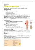
Nervous System Divisions
Central nervous system (CNS): includes the brain and spinal cord.
Peripheral nervous system (PNS): encompasses all other neural elements.
Brain Structure; the brain has layers:
Cerebrum: the outermost layer of the brain.
- The brain contains approximately 86 to 87 billion neurons, most of which are located
in the cerebellum.
Cerebral cortex: a key component of the cerebrum.
Neurons feature axons and dendrites to facilitate communication.
• Axons: long extensions that may have collaterals to connect with other neurons.
• Dendrites: extensively branched to communicate with other neurons.
o The brain is a complex neural network with both long-distance and short
connections.
Brain Surface Features
• Gyri: ridges on the brain's surface.
• Sulci: grooves on the brain's surface.
Brain Lobes
Comprised of four lobes: frontal, parietal, temporal, and occipital.
• Frontal lobe: critical for higher cognitive
functions and is the largest lobe.
• Interconnectivity between the lobes is crucial
for proper brain function.
1
,Psychopharmacology Hoorcolleges
Specific Brain Areas
• Prefrontal cortex: a very advanced area, significant for
higher order functions (the most human part of the brain).
• Hippocampus: an older part of the cerebral cortex,
considered to be the archicortex in contrast to the neo-cortex.
o Neo-cortex: also found in other mammal brains.
• Amygdala: vital for the expression of emotions.
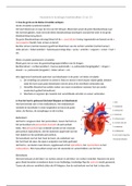
• Striatum and thalamus: involved in relay and processing
functions (can see in image a large band of white matter go
through it.
• Thalamus: a major relay station connecting different brain areas. Deeper in dicephalic
part of the brain. It connects different pathways in the brain.
• Hypothalamus: regulates homeostasis and autonomic control. It is the communication
between the conscious (higher order functions) and the more autonomic control of
many physiological part of the body (such as blood regulation).
• Occipital lobe: contains the primary visual cortex (V1), the starting point for visual
processing.
Matter Types
• Gray matter: consists of neuronal cell bodies.
• White matter: composed mainly of nerve fibers (axons).
The axons of grey matter are not heavily myelinated, unlike white matter, which contains a
high concentration of myelin.
Gray matter largely functions to receive information and regulate outgoing information, as it
contains the cell bodies of neurons. White matter, which is largely composed of axons, serves
to transmit signals to other regions of the brain, spinal cord, and body.
2
,Psychopharmacology Hoorcolleges
Biggest question in neuroscience: consciousness
Consciousness might be tied to structures within the brain, which are not fully understood and
vary among species.
- Know that it is a result of processing information from visual information (is the
starting point).
It is a top-down bottom-up process (both).
Animals have partially consciousness: they don’t have all of the structures we think are
important for generating consciousness.
Microstructure of the brain
Neuronal Cells: These are
the primary information
processors and transmitters
in the brain, with distinct
structures based on their
location:
• Pyramidal cells:
These are the
predominant cell type found in the cerebral cortex, characterized by their pyramid-
shaped cell bodies and long dendritic trees that are crucial for synaptic integration.
They play key roles in higher cognitive functions.
• Purkinje cells: Located in the cerebellum (area in the brain with the most neurons in
the brain), these cells have elaborated dendritic arbors that make them highly effective
at integrating synaptic inputs. The cerebellum, densely packed with neurons, functions
like a complex calculator, processing inputs to coordinate motor control and cognitive
functions.
Glial Cells: Supporting cells that are essential for maintaining the homeostasis of the brain's
environment and supporting the function of neurons (the function of your brain significantly
depends on glial cells):
• Microglial cells: These are the brain's resident immune cells. Upon activation by
infection or brain injury, microglial cells proliferate and mobilize to clear debris,
damaged cells, and pathogens through phagocytosis (they play a role in the defense
3
, Psychopharmacology Hoorcolleges
system: when the brain is infected, microglial cells will become activated, they can
help fight these pathogens and can help the brain repair itself). They are characterized
by small bodies and fine, branching extensions that probe the brain environment for
signs of disturbance.
o they remove components of damaged cells. They attack pathogens e.g., when
there is a viral cell in the brain tissue, or when there is a bacterial cell.
• Astrocytes: They regulate blood flow to active brain regions, enhancing the delivery
of oxygen and nutrients (take care that the very active parts of the brain get proper
oxygen).
o Astrocytes manage the clearance and recycling of neurotransmitters involved
in brain signaling (removing the excess of neurotransmitter compound).
o They provide metabolic support to neurons and are integral to the formation of
the blood-brain barrier, which protects the brain from harmful substances in the
bloodstream (provide the chemicals that neurons need to synthesize messenger
molecules; provide the interface between the neurons and blood supply).
§ These astrocytes play an important in that particular interface, they
sense activities in nerve cells and communicate with blood vessels in
your brain tissue to provide you with the necessary supply.
o In functional MRI (fMRI), these cells are crucial because they help visualize
brain activity by regulating blood flow in response to neuronal activity.
Ependymal cells: group of support cells – they don’t play such a functional roll, because they
mainly lie in the cavities and the large bodies in the brain, the ventricles (large gaps in the
brain).
Oligodendrocytes: These cells are vital for forming the myelin sheath around axons in the
CNS. Myelin is a fatty layer that insulates axons, allowing for the rapid transmission of
electrical signals. This insulation is crucial for the efficient functioning of the nervous system.
Nodes of Ranvier: These are small gaps in the myelin sheath where axonal membranes are
exposed. Electrical impulses jump from node to node in a
process known as saltatory conduction, which greatly
increases the speed of neural transmission.
4
Les 1: Neural signaling and neuromodulation
Nervous System Divisions
Central nervous system (CNS): includes the brain and spinal cord.
Peripheral nervous system (PNS): encompasses all other neural elements.
Brain Structure; the brain has layers:
Cerebrum: the outermost layer of the brain.
- The brain contains approximately 86 to 87 billion neurons, most of which are located
in the cerebellum.
Cerebral cortex: a key component of the cerebrum.
Neurons feature axons and dendrites to facilitate communication.
• Axons: long extensions that may have collaterals to connect with other neurons.
• Dendrites: extensively branched to communicate with other neurons.
o The brain is a complex neural network with both long-distance and short
connections.
Brain Surface Features
• Gyri: ridges on the brain's surface.
• Sulci: grooves on the brain's surface.
Brain Lobes
Comprised of four lobes: frontal, parietal, temporal, and occipital.
• Frontal lobe: critical for higher cognitive
functions and is the largest lobe.
• Interconnectivity between the lobes is crucial
for proper brain function.
1
,Psychopharmacology Hoorcolleges
Specific Brain Areas
• Prefrontal cortex: a very advanced area, significant for
higher order functions (the most human part of the brain).
• Hippocampus: an older part of the cerebral cortex,
considered to be the archicortex in contrast to the neo-cortex.
o Neo-cortex: also found in other mammal brains.
• Amygdala: vital for the expression of emotions.
• Striatum and thalamus: involved in relay and processing
functions (can see in image a large band of white matter go
through it.
• Thalamus: a major relay station connecting different brain areas. Deeper in dicephalic
part of the brain. It connects different pathways in the brain.
• Hypothalamus: regulates homeostasis and autonomic control. It is the communication
between the conscious (higher order functions) and the more autonomic control of
many physiological part of the body (such as blood regulation).
• Occipital lobe: contains the primary visual cortex (V1), the starting point for visual
processing.
Matter Types
• Gray matter: consists of neuronal cell bodies.
• White matter: composed mainly of nerve fibers (axons).
The axons of grey matter are not heavily myelinated, unlike white matter, which contains a
high concentration of myelin.
Gray matter largely functions to receive information and regulate outgoing information, as it
contains the cell bodies of neurons. White matter, which is largely composed of axons, serves
to transmit signals to other regions of the brain, spinal cord, and body.
2
,Psychopharmacology Hoorcolleges
Biggest question in neuroscience: consciousness
Consciousness might be tied to structures within the brain, which are not fully understood and
vary among species.
- Know that it is a result of processing information from visual information (is the
starting point).
It is a top-down bottom-up process (both).
Animals have partially consciousness: they don’t have all of the structures we think are
important for generating consciousness.
Microstructure of the brain
Neuronal Cells: These are
the primary information
processors and transmitters
in the brain, with distinct
structures based on their
location:
• Pyramidal cells:
These are the
predominant cell type found in the cerebral cortex, characterized by their pyramid-
shaped cell bodies and long dendritic trees that are crucial for synaptic integration.
They play key roles in higher cognitive functions.
• Purkinje cells: Located in the cerebellum (area in the brain with the most neurons in
the brain), these cells have elaborated dendritic arbors that make them highly effective
at integrating synaptic inputs. The cerebellum, densely packed with neurons, functions
like a complex calculator, processing inputs to coordinate motor control and cognitive
functions.
Glial Cells: Supporting cells that are essential for maintaining the homeostasis of the brain's
environment and supporting the function of neurons (the function of your brain significantly
depends on glial cells):
• Microglial cells: These are the brain's resident immune cells. Upon activation by
infection or brain injury, microglial cells proliferate and mobilize to clear debris,
damaged cells, and pathogens through phagocytosis (they play a role in the defense
3
, Psychopharmacology Hoorcolleges
system: when the brain is infected, microglial cells will become activated, they can
help fight these pathogens and can help the brain repair itself). They are characterized
by small bodies and fine, branching extensions that probe the brain environment for
signs of disturbance.
o they remove components of damaged cells. They attack pathogens e.g., when
there is a viral cell in the brain tissue, or when there is a bacterial cell.
• Astrocytes: They regulate blood flow to active brain regions, enhancing the delivery
of oxygen and nutrients (take care that the very active parts of the brain get proper
oxygen).
o Astrocytes manage the clearance and recycling of neurotransmitters involved
in brain signaling (removing the excess of neurotransmitter compound).
o They provide metabolic support to neurons and are integral to the formation of
the blood-brain barrier, which protects the brain from harmful substances in the
bloodstream (provide the chemicals that neurons need to synthesize messenger
molecules; provide the interface between the neurons and blood supply).
§ These astrocytes play an important in that particular interface, they
sense activities in nerve cells and communicate with blood vessels in
your brain tissue to provide you with the necessary supply.
o In functional MRI (fMRI), these cells are crucial because they help visualize
brain activity by regulating blood flow in response to neuronal activity.
Ependymal cells: group of support cells – they don’t play such a functional roll, because they
mainly lie in the cavities and the large bodies in the brain, the ventricles (large gaps in the
brain).
Oligodendrocytes: These cells are vital for forming the myelin sheath around axons in the
CNS. Myelin is a fatty layer that insulates axons, allowing for the rapid transmission of
electrical signals. This insulation is crucial for the efficient functioning of the nervous system.
Nodes of Ranvier: These are small gaps in the myelin sheath where axonal membranes are
exposed. Electrical impulses jump from node to node in a
process known as saltatory conduction, which greatly
increases the speed of neural transmission.
4