
Neuroscience – The senses
The 5 senses: touch, vision, smell, taste and
hearing
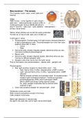
Vision
Optic nerves – come together in optic chiasm –
optic tract -- lateral geniculate nucleus (LGN) or
medial geniculate nucleus (MGM) (Brodman’s
area 41 and 42) – optic tract – optic radiation – diencephalon --
primary visual cortex
Retina: where photons are turned into action potentials.
Formed out of neural tube; optic cub is folded out
5 cell types in retina:
• Photoreceptors: Furthest away from light source, because detection
of light is damaging for cells. The photoreceptor turn over their outer
segment discs with 12 days.
o Cones
o Rods
• Horizontal cells; already integrate signals, determine what you see.
Connects to different photoreceptors
• Bipolar cells
• Amacrine cells; already integrate signals, determine what you see.
Connect to different ganglion cells
• Ganglion cells (their axons form the optic nerve)
Flow of information from photoreceptors – bipolar cells – ganglion cell
Photoreceptors:
• Rod: light sensitive receptor for black/white vision – Tall
o Dark: Na/Ca influx, K efflux, depolarization. cGMP is active
o Light: reduced Na/Ca influx, K efflux, hyperpolarization. cGMP
reduced
Retinal accepts a photon and activates opsin. G-coupled protein
receptor opsin activated transducin. Transducin activates
phosphodiesterase (PDE) which hydrolyses cGMP.
When cGMP level drops Ca/Na channel closes.
So, opsin receptor inactivates Na/Ca channel when
exposed to light.
• Cone: less sensitive receptor for coloured light – short
Distribution cones and rods:
Rods: present everywhere except in the fovea
Cods: present in the fovea
Better light detection in macula lutea
No sight in the optic disc (exit of optic nerve) (blind spot)
, Projections from photoreceptor:
Rods: 15-30 rods project on 1 bipolar cell – large receptive
field – high sensitivity
Rods in fovea project only to 1 bipolar cell – small receptive
field – low sensitivity
Cones: 1 rod project on bipolar cell – small receptive – low
sensitivity
Cone releases glutamate from its axon. Glutamate is filled
out where there is no light (dark).
2 types of bipolar cells to which they can project:
• OFF-center bipolar cell, hyperpolarized
• ON-center bipolar cell, depolarized.
Going to OFF-center?
• When there is light: less glutamate release, less
AMPA receptor (inotropic receptor). These will
hyperpolarize. From OFF-bipolar cell to OFF-center
ganglion cell also has AMPA, kainate, NMDA
receptor. These become inactive
• When there is NO light: glutamate release bind to
AMPA, kainate receptor. These will depolarize. From
OFF-bipolar cell to OFF-center ganglion cell also has
AMPA, kainate, NMDA receptor. These become
active
Going to ON-center?
• When there is light: less glutamate release bind to mGluR6 receptor. These
will depolarize. From OFF-bipolar cell to OFF-center ganglion cell also has
AMPA, kainate, NMDA receptor. These become active
• When there is NO light: glutamate release bind to mGluR6 receptor
(metabotropic receptor). These will hyperpolarize. From OFF-bipolar cell to
OFF-center ganglion cell also has AMPA, kainate, NMDA receptor. These
become inactive
Surround suppression by horizontal cells:
Horizontal cell integrates signals.
Hyperpolarization of surrounding cones by light results in hyperpolarization of
the connecting horizontal cells. These horizontal cells project on the central
cone and reduce their suppressive signals, leading to depolarization of the
central cone. This produces a strongly focused signal in the center receptive
field.
Optogenetics:
Biological technique in which light is used to control cells in living tissue, usually
neurons, that have been genetically modified to express light-sensitive ion channels.

