
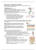
Neuroscience – Somatosensory sensation
Somatosensory system registers; touch (one of the 5 senses), temperature, pain,
pressure, vibration, limb position, itch
Somatosensory cortex
Located dorsal of the central sulcus.
Cortex contains a somatotropic maps; representation of the body in
separated regions. Some somatotropic maps larger compared to
relative size of the body part (thumb, lower lip).
Homonculus: reconstruction the body based on the somatotropic maps
in the somatosensory and motor cortex.
The cortex receives input from 2 type of fibers:
1. Mechanosensory afferent fiber; active by touch and
pressure. Detect stimulation using mechanoreceptors
2. Pain and temperature afferent fiber (nocireceptive
afferent); active by pain and temperature. Detect
stimulation using free nerve endings.
Both come from skin, both have cell bodies in dorsal root ganglion, both project in the
dorsal part of the spinal cord. The track of these fibers is different, level of crossing
over is different
Mechanoreceptors for touch/pressure/vibration found in;
Different mechanoreceptors detect a change in pressure in the
skin --> touch
Change of phospholipid pressure in the membrane leaflet
operates opening/closing of ion channels (conductive for Na).
When lateral position of membrane is stretched, ion channel
opens.
Receptive field: portion of sensory space that can elicit
(uitlokken) neuronal responses when stimulated
• Large receptive field – imprecision – high sensitivity, need not much
stimulation to evoke action potential – small in somatotropic map
• Small receptive field – precision – low sensitivity, need much stimulation to
evoke action potential – big in somatotropic map
Receptive field differs in size over the body; fingers small field, thigh big field.
Different mechanoreceptors in the skin
• Merkel cells; small receptive field (most sensitive one) –
information from superficial skin
• Meissner corpuscle; small receptive field – information from skin
• Ruffini corpuscle; large receptive field – detects stretching (finger
go from left to right or the other way around – information form
muscle
• Pacinian corpuscle; large receptive field– detects general
pressure – information from skin
, These mechanosensory afferent fibers detect:
• Change stimulation (rapid adapting); report that the stimulation
is on the skin and when the stimulation is released from the
skin. When is there a change?
• Persistent stimulation (slow adapting); report when the
stimulation is present. Duration of the stimulation?
Spinal cord;
• Sensory information from lower body; lumbar
spinal cord.
o Fasciculus gracilis (medial) --> gracile
tract
• Sensory information from upper body; cervical
spinal cord
o Fasciculus cuneatus (lateral) -->
cuneate tract
In the lumbar spinal cord only the fasciculus gracilis
exists.
In the cervical spinal cord both the fasciculus gracilis
and fasciculus cuneatus exists
Medulla;
• Sensory information from lower body; gracile
nucleus (medial) to midline of medulla. Now
this one lateral (here decussation
(omdraaien))
• Sensory information from upper body; cuneate
nucleus (lateral) to midline of medulla. Now
this one medial (here decussation (omdraaien))
Pons:
• Sensory information from lower body; to medial lemniscus
• Sensory information from upper body; to medial lemniscus
• Sensory information from face; come in via trigeminal ganglion via principal
nucleus of trigeminal complex to medial lemniscus (here decussation
(omdraaien))
Midbrain;
• Sensory information from lower body; to medial
lemniscus
• Sensory information from upper body; to medial
lemniscus
• Sensory information from face; to trigeminal
lemniscus
Brain;
• Sensory information from lower body; to thalamus
(ventral posterior lateral nucleus) via capsula


