Week 1: Tumor Biology & Clinical Behavior
LECTURE 1: HISTOPATHOLOGY – TUMOR STAGING (W.J. Mooi) Monday, 29/10/2018
Cancer detection & diagnosis (identification & determination of cancer cell type):
a. Screening: chance finding in asymptomatic patient
b. Index complaint (symptoms appear: e.g. lump, etc set into motion the Dx cascade)
c. Phys Ex (symptoms +)
d. Radiologic investigation
e. Diagnostic nuclear medicine (radioactive trecer)
f. Clinical test: blood test, Dx microscopy/pathology (e.g. cytopathology exfo, FNA, histopathology of biopsy &
resection specimen)
Triple diagnosis of cancer: physical ex, histopathology, radiology
Exfoliative cytology
Exfoliative cytology (tissue brushing): looking at cells/group of cells outside the tissue context, may identify metastatic cells,
dysplastic cells, etc non-intact tissue sample; Sources: body cavities, body fluids, etc
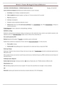
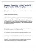
Fine needle aspiration/FNA
Method: palpation/USG-guided benefit: no anesthesia, quick, small needle
Needle biopsy enables the examination of tumor cells from intact environment i.e. in the actual site
Fine needle: cytology
Thick needle: histopathology (more representative for actual tumor tissue compared to FNA)
Endoscopic biopsy
Endoscope + blade for tissue excision (“biting” the tissue at lump site) disadvantage: should be anesthetized (performed in
OR/sterile room)
Other method of tissue procurement: punch biopsy (skin lesions), traditional biopsy (e.g. BMP), traditional excision biopsy for
superficial lesions
Pathologic tumor diagnosis
Routine light microscopy: staining procedures (HE, Giemsa, Papanicolau, etc) differentiation of different cells
Histochemical stains
IHC: identifying antigens present in tissue samples certain cells would bind more strongly to certain antibody
ISH
Molecular analysis: gene expression (microarray), mutation detection, CGH, LOH mapping
, Week 1: Tumor Biology & Clinical Behavior
Procedures of tissue sampling
Aim: determine the scope of disease in tissue sample & identify clinically relevant area of the biopsy sample, e.g. presence of
tumor cells in resection margins Not all sample area gets analyzed, adjust with the needs of clinicians
Example: FFPE Definition of FFPE: …?
Procedure: tissue taken out of patient transferred to pathology formaldehyde stabilization wash off, replace with
paraffin (dehydration of sample & further stabilization) cooling tissue block examination in microtome slicing off
the tissue rehydration staining of sample specimen analysis on microscope
Crosslink of molecules when treated with formaldehyde: …?
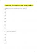
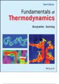
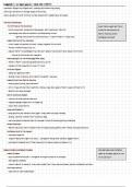
General histologic properties of cancer (“cardinal signs”)
Anaplasia (loss of differentiation) Pleomorphism of cells & nuclei genetic mutations
result in presence of variants that present as different
Presence of keratin accumulation/“keratin pearl” cellular/nuclear morphology (small/large nuclei, heavily
indicates squamous differentiation, “undifferentiated” stained/unstained)
region of cells w/ aberrant differentiation
)
Disordered cellular architecture Abnormal mitosis
Conditions of basement membrane, presence of normal Presence of mitotic figures (double nuclei, abnormal
cellular structures, invasion, etc separation of cells, abnormal daughter cells/”triaster”)
, Week 1: Tumor Biology & Clinical Behavior
Tumor grading
a. Cytological atypia c. Presence of necrosis
b. Mitotic activity d. Degree of resemblance to normal tissue
(differentiation)
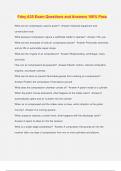
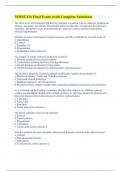
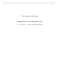
Invasive growth
Example: BCC cutaneous spread down the subcutaneous, heavy staining of nuclei (abnormal mitosis)
Types:
a. Lymphogenic
b. Hematogenic
c. Direct seeding (e.g. to adjacent organ, through body cavities)
Angioinvasion: presence of tumor cells inside blood/lymph vessels on histology sample most common route for metastasis
(e.g. loss of basement membrane underneath the vascular endothelium, rearranged stroma, tumor clones inside vessel)
Importance of pathological cancer classification
Tumors differ in growth rate, destructiveness for invasion, propensity for metastasis (regional, distant), response to Tx,
prognosis, family history, etc complex classification in order to determine the therapeutic options to employ for certain
patient
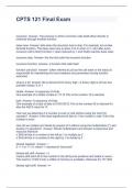
1. Determining the main groups of
malignant tumors: carcinoma,
sarcoma, hematopoietic tumor, germ
cell tumor/GCT, lymphoma, brain
tumor
2. Cancer grading (low-grade/ well-
differentiated, high-grade/poorly
differentiated, undifferentiated)
histopathological
3. Tumor spread/stage grouping (TNM
staging Tumor, lymph Node, distant
hematogenous Metastasis; TNM later
utilized for stage-grouping 0-4, details
see slide) clinical
LECTURE 2: LAB TECHNIQUES/TOOLS (S. Cilessen) Monday, 29/10/2018
Molecular detection techniques: DNA, RNA, protein, cellular functional assay, animal model, molecular imaging
Always perform validation (compare the preferred technique with easier, more large-scale techniques e.g. PCR followed
by mRNA assay, IHC, etc)
, Week 1: Tumor Biology & Clinical Behavior
DNA Assay
a. NGS/MPS
Determine accurate order of nucleotides
(A/G/T/C) along chromosomes & genomes
analyzing mutated pathways, resistance
mechanism, etc
Advantage: can be applied for many different
samples with low quantity of sample
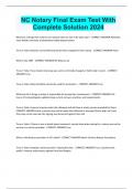
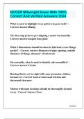
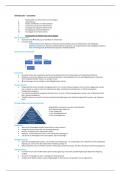
b. Array CGH c. Methylated DNA immunoprecipitation (MEDIP)
Analyze CNV (gains/loss) Denature DNA label w/ antibody pattern formation by
antibody immunoprecipitation enriched Me-DNA
Extract DNA + digest label tumor (Cy5) &
production high throughput sequencing; can also use the
reference (Cy3) DNA combine equal amounts of
denatured DNA for array hybridization
DNA hybridization image analysis in software
(spots)
LECTURE 1: HISTOPATHOLOGY – TUMOR STAGING (W.J. Mooi) Monday, 29/10/2018
Cancer detection & diagnosis (identification & determination of cancer cell type):
a. Screening: chance finding in asymptomatic patient
b. Index complaint (symptoms appear: e.g. lump, etc set into motion the Dx cascade)
c. Phys Ex (symptoms +)
d. Radiologic investigation
e. Diagnostic nuclear medicine (radioactive trecer)
f. Clinical test: blood test, Dx microscopy/pathology (e.g. cytopathology exfo, FNA, histopathology of biopsy &
resection specimen)
Triple diagnosis of cancer: physical ex, histopathology, radiology
Exfoliative cytology
Exfoliative cytology (tissue brushing): looking at cells/group of cells outside the tissue context, may identify metastatic cells,
dysplastic cells, etc non-intact tissue sample; Sources: body cavities, body fluids, etc
Fine needle aspiration/FNA
Method: palpation/USG-guided benefit: no anesthesia, quick, small needle
Needle biopsy enables the examination of tumor cells from intact environment i.e. in the actual site
Fine needle: cytology
Thick needle: histopathology (more representative for actual tumor tissue compared to FNA)
Endoscopic biopsy
Endoscope + blade for tissue excision (“biting” the tissue at lump site) disadvantage: should be anesthetized (performed in
OR/sterile room)
Other method of tissue procurement: punch biopsy (skin lesions), traditional biopsy (e.g. BMP), traditional excision biopsy for
superficial lesions
Pathologic tumor diagnosis
Routine light microscopy: staining procedures (HE, Giemsa, Papanicolau, etc) differentiation of different cells
Histochemical stains
IHC: identifying antigens present in tissue samples certain cells would bind more strongly to certain antibody
ISH
Molecular analysis: gene expression (microarray), mutation detection, CGH, LOH mapping
, Week 1: Tumor Biology & Clinical Behavior
Procedures of tissue sampling
Aim: determine the scope of disease in tissue sample & identify clinically relevant area of the biopsy sample, e.g. presence of
tumor cells in resection margins Not all sample area gets analyzed, adjust with the needs of clinicians
Example: FFPE Definition of FFPE: …?
Procedure: tissue taken out of patient transferred to pathology formaldehyde stabilization wash off, replace with
paraffin (dehydration of sample & further stabilization) cooling tissue block examination in microtome slicing off
the tissue rehydration staining of sample specimen analysis on microscope
Crosslink of molecules when treated with formaldehyde: …?
General histologic properties of cancer (“cardinal signs”)
Anaplasia (loss of differentiation) Pleomorphism of cells & nuclei genetic mutations
result in presence of variants that present as different
Presence of keratin accumulation/“keratin pearl” cellular/nuclear morphology (small/large nuclei, heavily
indicates squamous differentiation, “undifferentiated” stained/unstained)
region of cells w/ aberrant differentiation
)
Disordered cellular architecture Abnormal mitosis
Conditions of basement membrane, presence of normal Presence of mitotic figures (double nuclei, abnormal
cellular structures, invasion, etc separation of cells, abnormal daughter cells/”triaster”)
, Week 1: Tumor Biology & Clinical Behavior
Tumor grading
a. Cytological atypia c. Presence of necrosis
b. Mitotic activity d. Degree of resemblance to normal tissue
(differentiation)
Invasive growth
Example: BCC cutaneous spread down the subcutaneous, heavy staining of nuclei (abnormal mitosis)
Types:
a. Lymphogenic
b. Hematogenic
c. Direct seeding (e.g. to adjacent organ, through body cavities)
Angioinvasion: presence of tumor cells inside blood/lymph vessels on histology sample most common route for metastasis
(e.g. loss of basement membrane underneath the vascular endothelium, rearranged stroma, tumor clones inside vessel)
Importance of pathological cancer classification
Tumors differ in growth rate, destructiveness for invasion, propensity for metastasis (regional, distant), response to Tx,
prognosis, family history, etc complex classification in order to determine the therapeutic options to employ for certain
patient
1. Determining the main groups of
malignant tumors: carcinoma,
sarcoma, hematopoietic tumor, germ
cell tumor/GCT, lymphoma, brain
tumor
2. Cancer grading (low-grade/ well-
differentiated, high-grade/poorly
differentiated, undifferentiated)
histopathological
3. Tumor spread/stage grouping (TNM
staging Tumor, lymph Node, distant
hematogenous Metastasis; TNM later
utilized for stage-grouping 0-4, details
see slide) clinical
LECTURE 2: LAB TECHNIQUES/TOOLS (S. Cilessen) Monday, 29/10/2018
Molecular detection techniques: DNA, RNA, protein, cellular functional assay, animal model, molecular imaging
Always perform validation (compare the preferred technique with easier, more large-scale techniques e.g. PCR followed
by mRNA assay, IHC, etc)
, Week 1: Tumor Biology & Clinical Behavior
DNA Assay
a. NGS/MPS
Determine accurate order of nucleotides
(A/G/T/C) along chromosomes & genomes
analyzing mutated pathways, resistance
mechanism, etc
Advantage: can be applied for many different
samples with low quantity of sample
b. Array CGH c. Methylated DNA immunoprecipitation (MEDIP)
Analyze CNV (gains/loss) Denature DNA label w/ antibody pattern formation by
antibody immunoprecipitation enriched Me-DNA
Extract DNA + digest label tumor (Cy5) &
production high throughput sequencing; can also use the
reference (Cy3) DNA combine equal amounts of
denatured DNA for array hybridization
DNA hybridization image analysis in software
(spots)