
UCI BioSci D170, Williams Digestion
Reading: Chapter 23
What organs make up the alimentary canal?
The GI (gastrointestinal) tract is made up of the mouth, pharynx, esophagus, stomach,
small intestine, large intestine, and anus.
What organs / structures make up the accessory digestive organs?
Consist of the teeth, tongue, gallbladder, and large digestive glands (salivary glands,
liver, and pancreas).
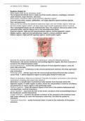
What are the four planes that divide the abdominal region into nine smaller regions? What are
the nine smaller regions called? List each major organ and the quadrant(s) where it is found.
The 2 parasagittal planes are the midclavicular lines; the superior transverse plane is the
subcostal plane, and the inferior one is the transtubercular plane.
Superior regions: right and left hypochondriac regions, central epigastric region.
Middle regions: right and left lateral/lumbar regions, central umbilical region.
Inferior regions: right and left inguinal regions, central pubic region.
Describe the structure and function of the peritoneum, noting the following structures:
Peritoneum is the serous membrane surrounding digestive organs; the most extensive is
located in the abdominopelvic cavity.
Visceral peritoneum – covers the external surfaces of most digestive organs. Lines the
inner tube externally.
Parietal peritoneum – continuous w/ the visceral peritoneum and lines the body wall (outer
tube internally).
Peritoneal cavity – slit-like space b/w the visceral and parietal peritoneum that contains
cerour fluid. -> allows digestive organs to easily glide along the body wall.
What is a mesentery? What are its functions? Describe the location and function of the following
mesenteries, including what organs they attach to:
Mesentery is a double-layer of peritoneum that extends from the body wall to the
digestive organs. It holds organs in place, stores fat, and provides a route for circulatory
vessels/nerves to reach organs in the peritoneal cavity.
Falciform ligament – binds the anterior aspect of the liver to the anterior abdominal wall
and diaphragm. (ventral mesentery)
Lesser omentum – runs from the liver to the lesser curvature of the stomach/beginning of
the duodenum. (ventral mesentery)
Greater omentum – connects the greater curvature of the stomach to the posterior body
wall; loops inferiorly to cover the transverse colon first. Can limit the spread of
infections.
Transverse mesocolon – nearly-horizontal sheet is fused to the underside of the greater
omentum.
, UCI BioSci D170, Williams Digestion
Sigmoid mesocolon – connects the sigmoid colon to the posterior pelvic wall.
Which organs lack mesenteries? (secondary retroperitoneal organs)
Pancreas, duodenum, ascending and descending colon, and rectum.
What are the six functions of the digestive system?
Ingestion, propulsion (swallowing, peristalsis), mechanical breakdown (chewing,
stomach churning, segmentation), digestion (breaking down food into their chemical
building blocks via enzymes), absorption, and defecation.
Describe the general microscopic anatomy of the alimentary canal, noting the following layers
and structures:
The alimentary canal from the esophagus to the anus has 4 tissue layers (NOT mouth).
Internal->external...
Mucosa – the innermost layer containing 3 sublayers: a lining epithelium (borders the
lumen), a lamina propria (vascularized CT that nourishes the lining, absorbs digested
Reading: Chapter 23
What organs make up the alimentary canal?
The GI (gastrointestinal) tract is made up of the mouth, pharynx, esophagus, stomach,
small intestine, large intestine, and anus.
What organs / structures make up the accessory digestive organs?
Consist of the teeth, tongue, gallbladder, and large digestive glands (salivary glands,
liver, and pancreas).
What are the four planes that divide the abdominal region into nine smaller regions? What are
the nine smaller regions called? List each major organ and the quadrant(s) where it is found.
The 2 parasagittal planes are the midclavicular lines; the superior transverse plane is the
subcostal plane, and the inferior one is the transtubercular plane.
Superior regions: right and left hypochondriac regions, central epigastric region.
Middle regions: right and left lateral/lumbar regions, central umbilical region.
Inferior regions: right and left inguinal regions, central pubic region.
Describe the structure and function of the peritoneum, noting the following structures:
Peritoneum is the serous membrane surrounding digestive organs; the most extensive is
located in the abdominopelvic cavity.
Visceral peritoneum – covers the external surfaces of most digestive organs. Lines the
inner tube externally.
Parietal peritoneum – continuous w/ the visceral peritoneum and lines the body wall (outer
tube internally).
Peritoneal cavity – slit-like space b/w the visceral and parietal peritoneum that contains
cerour fluid. -> allows digestive organs to easily glide along the body wall.
What is a mesentery? What are its functions? Describe the location and function of the following
mesenteries, including what organs they attach to:
Mesentery is a double-layer of peritoneum that extends from the body wall to the
digestive organs. It holds organs in place, stores fat, and provides a route for circulatory
vessels/nerves to reach organs in the peritoneal cavity.
Falciform ligament – binds the anterior aspect of the liver to the anterior abdominal wall
and diaphragm. (ventral mesentery)
Lesser omentum – runs from the liver to the lesser curvature of the stomach/beginning of
the duodenum. (ventral mesentery)
Greater omentum – connects the greater curvature of the stomach to the posterior body
wall; loops inferiorly to cover the transverse colon first. Can limit the spread of
infections.
Transverse mesocolon – nearly-horizontal sheet is fused to the underside of the greater
omentum.
, UCI BioSci D170, Williams Digestion
Sigmoid mesocolon – connects the sigmoid colon to the posterior pelvic wall.
Which organs lack mesenteries? (secondary retroperitoneal organs)
Pancreas, duodenum, ascending and descending colon, and rectum.
What are the six functions of the digestive system?
Ingestion, propulsion (swallowing, peristalsis), mechanical breakdown (chewing,
stomach churning, segmentation), digestion (breaking down food into their chemical
building blocks via enzymes), absorption, and defecation.
Describe the general microscopic anatomy of the alimentary canal, noting the following layers
and structures:
The alimentary canal from the esophagus to the anus has 4 tissue layers (NOT mouth).
Internal->external...
Mucosa – the innermost layer containing 3 sublayers: a lining epithelium (borders the
lumen), a lamina propria (vascularized CT that nourishes the lining, absorbs digested

