
Cell Structure and Function (ch. 3, lec. 3)
Tuesday, October 6, 2020 8:36 PM
Cell membrane
• Prokaryotic cells do not have organelles enclosed in membranes, instead organized in distinct
locations.
• Cell membrane: selectively permeable barrier that separates cellular components from external
environment.
○ Membranes have equal volumes of phospholipids (glycerol w/ ester links to 2 fatty acids and a
phosphoryl head group) and proteins.
○ Leaflets: 2 layers of phospholipids in the bilayer.
• Phospholipids vary by phosphoryl head groups and fatty acid side chains. Ex. Cardiolipin: double
phospholipid linked by a glycerol. Its conc INCs under cell stress; localizes to cell poles (binds to
certain environmental stress proteins/like protein that transfers osmo-protectants).
○ Fatty acid chains may be saturated, unsaturated, or polyunsaturated.
• Membranes also include planar molecules that fill gaps b/w hydrocarbon chains.
○ In eukaryotes, reinforcing agents are sterols (ie cholesterol).
○ In bacteria, agents are hopanoids.
• Archaea have the most extreme variations in phospholipid side chains.
○ Ether links b/w glycerol and fatty acids are diethers or tetraethers. Hydrocarbon chains are
branched terpenoids (can cyclize to form cyclopentane).
○ Live in harsh environments. -> Need diversity in their membrane structure to survive in
extremes.
○ *Archaea have ETHER linkages while bacteria have ester.
Membrane proteins
• Phospholipid membranes have hydrophilic heads on outside and hydrophobic fatty acids facing
inside of cell.
○ Membrane proteins are able to float inside said structures and transport thru the membrane.
○ They also have hydrophilic and hydrophobic regions, allowing them to be inserted into the cell
membrane.
• Membrane proteins serve many functions: structural support, detection of environmental signals,
secretion of virulence factors and communication signals, ion transport and energy storage.
Cell wall
• Confers shape and rigidity; helps withstand turgor pressure.
• Cell wall is a single molecule. The sacculus, bacterial cell wall, consists of a single interlinked
molecule.
• Most bacterial walls are made of peptidoglycan (murein)-- long polymers of repeating
disaccharides bound to a 4-6 amino acid peptide. Peptides can form cross-bridges connecting
parallel glycan strands (b/w 3rd and 4th aa).
• Peptidoglycan forms a helical strand. -> proteins up to 50kDa can pass through!
• Peptidoglycan is unique to bacteria. -> Enzymes responsible for its biosynthesis make great targets
for antibiotics. Ex. Penicillin inhibits the transpeptidase that cross-links the peptides.
3.3: The Envelope and Cytoskeleton
• For many prokaryotic species, the cell envelope includes at least one layer of support (ie cell wall).
• Envelope composition defines whether a bacteria is: (assume all components are in cell wall)
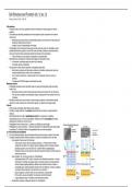
○ Gram positive: thick cell wall. Ex Firmicutes
• Teichoic acids: anchor cell wall to plasma membrane. -> Protect cell from harmful
substances, important for ion uptake, help their pathogens bind to host tissue. Made of
phosphate, glycerol, and a side chain (R). Covalently linked to peptidoglycans.
• Periplasmic space: narrow space b/w cell wall and plasma membrane; contains structural
proteins/exoenzymes.
○ Gram negative: thin, yet strong cell wall. Ex. Proteobacteria
• Lipopolysaccharides (LPS): Lipid A component is an inflammatory endotoxin -> induces
inflammation. Contributes to negative charge on bacterial surface (core polysaccharide
contains charged sugars/phosphate), stabilize outer membrane structure, creates a
permeability layer, protects pathogenic bacteria from host defenses. Contains a repeating
unit (elicits immune response), outer and inner core, and lipid A.
• Braun's lipoprotein: connects murein w/ outer membrane, most abundant.
• Porin proteins: assure permeability. Tube-shaped, water-filled channels.
• Large periplasmic space w/ hydrolytic enzymes/transport proteins.
○ Mycobacteria: complex, multilayered cell wall. Ex. Actinobacteria
Tuesday, October 6, 2020 8:36 PM
Cell membrane
• Prokaryotic cells do not have organelles enclosed in membranes, instead organized in distinct
locations.
• Cell membrane: selectively permeable barrier that separates cellular components from external
environment.
○ Membranes have equal volumes of phospholipids (glycerol w/ ester links to 2 fatty acids and a
phosphoryl head group) and proteins.
○ Leaflets: 2 layers of phospholipids in the bilayer.
• Phospholipids vary by phosphoryl head groups and fatty acid side chains. Ex. Cardiolipin: double
phospholipid linked by a glycerol. Its conc INCs under cell stress; localizes to cell poles (binds to
certain environmental stress proteins/like protein that transfers osmo-protectants).
○ Fatty acid chains may be saturated, unsaturated, or polyunsaturated.
• Membranes also include planar molecules that fill gaps b/w hydrocarbon chains.
○ In eukaryotes, reinforcing agents are sterols (ie cholesterol).
○ In bacteria, agents are hopanoids.
• Archaea have the most extreme variations in phospholipid side chains.
○ Ether links b/w glycerol and fatty acids are diethers or tetraethers. Hydrocarbon chains are
branched terpenoids (can cyclize to form cyclopentane).
○ Live in harsh environments. -> Need diversity in their membrane structure to survive in
extremes.
○ *Archaea have ETHER linkages while bacteria have ester.
Membrane proteins
• Phospholipid membranes have hydrophilic heads on outside and hydrophobic fatty acids facing
inside of cell.
○ Membrane proteins are able to float inside said structures and transport thru the membrane.
○ They also have hydrophilic and hydrophobic regions, allowing them to be inserted into the cell
membrane.
• Membrane proteins serve many functions: structural support, detection of environmental signals,
secretion of virulence factors and communication signals, ion transport and energy storage.
Cell wall
• Confers shape and rigidity; helps withstand turgor pressure.
• Cell wall is a single molecule. The sacculus, bacterial cell wall, consists of a single interlinked
molecule.
• Most bacterial walls are made of peptidoglycan (murein)-- long polymers of repeating
disaccharides bound to a 4-6 amino acid peptide. Peptides can form cross-bridges connecting
parallel glycan strands (b/w 3rd and 4th aa).
• Peptidoglycan forms a helical strand. -> proteins up to 50kDa can pass through!
• Peptidoglycan is unique to bacteria. -> Enzymes responsible for its biosynthesis make great targets
for antibiotics. Ex. Penicillin inhibits the transpeptidase that cross-links the peptides.
3.3: The Envelope and Cytoskeleton
• For many prokaryotic species, the cell envelope includes at least one layer of support (ie cell wall).
• Envelope composition defines whether a bacteria is: (assume all components are in cell wall)
○ Gram positive: thick cell wall. Ex Firmicutes
• Teichoic acids: anchor cell wall to plasma membrane. -> Protect cell from harmful
substances, important for ion uptake, help their pathogens bind to host tissue. Made of
phosphate, glycerol, and a side chain (R). Covalently linked to peptidoglycans.
• Periplasmic space: narrow space b/w cell wall and plasma membrane; contains structural
proteins/exoenzymes.
○ Gram negative: thin, yet strong cell wall. Ex. Proteobacteria
• Lipopolysaccharides (LPS): Lipid A component is an inflammatory endotoxin -> induces
inflammation. Contributes to negative charge on bacterial surface (core polysaccharide
contains charged sugars/phosphate), stabilize outer membrane structure, creates a
permeability layer, protects pathogenic bacteria from host defenses. Contains a repeating
unit (elicits immune response), outer and inner core, and lipid A.
• Braun's lipoprotein: connects murein w/ outer membrane, most abundant.
• Porin proteins: assure permeability. Tube-shaped, water-filled channels.
• Large periplasmic space w/ hydrolytic enzymes/transport proteins.
○ Mycobacteria: complex, multilayered cell wall. Ex. Actinobacteria

