

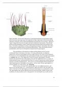
Figure 22.6 (left). The hornwort Phaeoceros, showing gametophyte and sporophyte stages.
Mature hornwort sporangia split into longitudinal strips, beginning at the tip and opening
downward. Rhizoids, which anchor gametophytes to the ground, are not shown. Figure 22.7
(right). A longitudinal section through a hornwort (Anthoceros) sporophyte. Only the mid-
portion of the sporophyte is shown, omitting the tip of the sporangium and rhizoids at the
base. The tubular sporophyte is mainly an elongated sporangium. Sporophyte cells within
the sporangium undergo meiosis to produce haploid spores. Other cells produce twisted
pseudoelaters, which assist in the ejection of spores from the sporangium.
The sporophyte of hornworts is unique among plants in that it grows
continually from a meristem at its base. It is long and pointed, creating the
appearance of horns protruding from the thallus (hence, the name of the division).
It consists of a foot embedded in the gametophyte thallus and an upright sporangium
or capsule (Fig. 22.7). The epidermis of some hornwort sporophytes contains
stomata but generally lacks chloroplasts; beneath the epidermis there is a layer of
chlorenchyma tissue, and beneath this there is a mass of sporocytes, which undergo
meiosis to produce haploid spores. The spores are intermingled with pseudoelaters,
which help separate and disperse the spores. In the center of the sporangium is a
central cylinder of sterile tissue, the columella.
When mature, the tip of the capsule splits into two valves (sections), and
spores are released (Fig. 22.7). A meristematic region just above the foot adds new
cells to the base of the sporangium so that more sporocytes are continually created.
The sporangium thus grows upward from the base much like a blade of grass and
continues to release spores over a long period. Its total height may reach several
10
,centimeters. If the spores land in a suitable environment, they undergo mitosis and
produce new gametophytes.
22.4 LIVERWORTS
Approximately 9,000 species of plants comprise the group known as liverworts, or
hepatics (derived from the Greek word meaning "liver"). The name is old, having
been recorded in medieval manuscripts as early as the ninth century A.D. It was
probably applied to these plants because of their fancied resemblance to the liver
and the belief that plants could cure diseases of the organs that they resembled. A
prescription for a liver complaint in the sixteenth century called for "liverworts
soaked in wine."
As is the case with all bryophytes, the liverwort gametophyte is the more
prominent phase of the life cycle. Most grow in moist, shady habitats. Those in
temperate regions usually grow as a green ribbon or heart-shaped band of tissue
(Fig. 22.8a). These thalli can resemble hornworts. The thallus is held to the surface
of the damp soil by single-celled rhizoids. Individual plants are small, ranging in size
from one to several centimeters across. However, colonies of liverworts can occupy
large areas.
Some liverworts are more elaborate, with distinct leaves and stems. The
leaves are blunt-tipped or lobed and are attached to the stems in two or three
overlapping rows (Fig. 22.8b). The gametophytes of liverworts produce an enormous
variety of volatile oils, which are stored in a unique single membrane-enclosed
organelle called an oil body. These oils give many liverworts a distinctive aroma,
and they may serve to prevent herbivory. Some of these compounds are promising
as antibiotics and anti-tumor agents.
a b
Figure 22.8. Liverworts.
(a) Riccia gametophytes, seen
from above. (b) A leafy liver-
wort, Porella, showing an
entire plant and details of the
stem and leaves.
11
, Liverworts produce relatively simple sporophytes that are hidden in folds in
the gametophyte tissue. As spores develop, special cells near the base of the
sporophyte elongate rapidly to form a stalk, called a seta, which pushes the
sporangium, or capsule, above the surface of the thallus. The capsule then splits into
four valves and releases all of its spores at one time. Special thickened cells inside
the capsules, called elaters, separate the spores and aid in spore dispersal. If the
spores land in a suitable environment, they undergo mitosis and produce one new
gametophyte plant per spore.
Our discussion of this group focuses on the genera Riccia and Marchantia, two
types of thalloid liverworts, and the genus Porella, a leafy liverwort. These three
examples represent the morphological variation of hepatic plants.
Some Liverworts Have Thalloid Bodies
Gametophytes of the thalloid (ribbon-shaped) liverworts usually grow Y-shaped
branches by a simple forking at the growing tip. In some species, a rosette of
branches is formed. About 15% of all liverwort species are thalloid, and of these
about two-thirds have simple undifferentiated thalli. The remaining third have more
complex thalli with distinct layers visible.
The gametophyte of
Marchantia is a good example of a
thalloid liverwort. It has a prominent
midrib, and the tips of the branches
are notched. A typical thallus is 1 to 2
cm across. The size and degree of
branching depend on growing
conditions. On the upper epidermis
are polygonal areas, each with a
conspicuous pore in the center (Fig.
22.9). These areas demarcate air
chambers below the pore, which bathe
chlorenchyma cells in air containing
carbon dioxide. The pores perform
the same function as stomata. The
lower cells of the thallus are colorless Figure 22.9. Cross section of a Marchantia
parenchyma, modified for thallus, showing the pore, air chamber,
carbohydrate storage. Rhizoids and epidermis, chlorenchyma, and
sheets of cells called scales project parenchyma storage tissue.
from the lower surface, increasing the
surface area in contact with the
substrate and anchoring the thallus.
Marchantia reproduces asexually in two ways: by fragmentation when older
parts of the thallus die, separating younger portions that then develop as separate
individuals; and by small clumps of tissue called gemmae (singular, gemma). These
clumps are produced in small gemmae cups that form on the upper surface of the
thallus (Fig. 22.10). When the gemmae are mature, raindrops can break them free of
12
Mature hornwort sporangia split into longitudinal strips, beginning at the tip and opening
downward. Rhizoids, which anchor gametophytes to the ground, are not shown. Figure 22.7
(right). A longitudinal section through a hornwort (Anthoceros) sporophyte. Only the mid-
portion of the sporophyte is shown, omitting the tip of the sporangium and rhizoids at the
base. The tubular sporophyte is mainly an elongated sporangium. Sporophyte cells within
the sporangium undergo meiosis to produce haploid spores. Other cells produce twisted
pseudoelaters, which assist in the ejection of spores from the sporangium.
The sporophyte of hornworts is unique among plants in that it grows
continually from a meristem at its base. It is long and pointed, creating the
appearance of horns protruding from the thallus (hence, the name of the division).
It consists of a foot embedded in the gametophyte thallus and an upright sporangium
or capsule (Fig. 22.7). The epidermis of some hornwort sporophytes contains
stomata but generally lacks chloroplasts; beneath the epidermis there is a layer of
chlorenchyma tissue, and beneath this there is a mass of sporocytes, which undergo
meiosis to produce haploid spores. The spores are intermingled with pseudoelaters,
which help separate and disperse the spores. In the center of the sporangium is a
central cylinder of sterile tissue, the columella.
When mature, the tip of the capsule splits into two valves (sections), and
spores are released (Fig. 22.7). A meristematic region just above the foot adds new
cells to the base of the sporangium so that more sporocytes are continually created.
The sporangium thus grows upward from the base much like a blade of grass and
continues to release spores over a long period. Its total height may reach several
10
,centimeters. If the spores land in a suitable environment, they undergo mitosis and
produce new gametophytes.
22.4 LIVERWORTS
Approximately 9,000 species of plants comprise the group known as liverworts, or
hepatics (derived from the Greek word meaning "liver"). The name is old, having
been recorded in medieval manuscripts as early as the ninth century A.D. It was
probably applied to these plants because of their fancied resemblance to the liver
and the belief that plants could cure diseases of the organs that they resembled. A
prescription for a liver complaint in the sixteenth century called for "liverworts
soaked in wine."
As is the case with all bryophytes, the liverwort gametophyte is the more
prominent phase of the life cycle. Most grow in moist, shady habitats. Those in
temperate regions usually grow as a green ribbon or heart-shaped band of tissue
(Fig. 22.8a). These thalli can resemble hornworts. The thallus is held to the surface
of the damp soil by single-celled rhizoids. Individual plants are small, ranging in size
from one to several centimeters across. However, colonies of liverworts can occupy
large areas.
Some liverworts are more elaborate, with distinct leaves and stems. The
leaves are blunt-tipped or lobed and are attached to the stems in two or three
overlapping rows (Fig. 22.8b). The gametophytes of liverworts produce an enormous
variety of volatile oils, which are stored in a unique single membrane-enclosed
organelle called an oil body. These oils give many liverworts a distinctive aroma,
and they may serve to prevent herbivory. Some of these compounds are promising
as antibiotics and anti-tumor agents.
a b
Figure 22.8. Liverworts.
(a) Riccia gametophytes, seen
from above. (b) A leafy liver-
wort, Porella, showing an
entire plant and details of the
stem and leaves.
11
, Liverworts produce relatively simple sporophytes that are hidden in folds in
the gametophyte tissue. As spores develop, special cells near the base of the
sporophyte elongate rapidly to form a stalk, called a seta, which pushes the
sporangium, or capsule, above the surface of the thallus. The capsule then splits into
four valves and releases all of its spores at one time. Special thickened cells inside
the capsules, called elaters, separate the spores and aid in spore dispersal. If the
spores land in a suitable environment, they undergo mitosis and produce one new
gametophyte plant per spore.
Our discussion of this group focuses on the genera Riccia and Marchantia, two
types of thalloid liverworts, and the genus Porella, a leafy liverwort. These three
examples represent the morphological variation of hepatic plants.
Some Liverworts Have Thalloid Bodies
Gametophytes of the thalloid (ribbon-shaped) liverworts usually grow Y-shaped
branches by a simple forking at the growing tip. In some species, a rosette of
branches is formed. About 15% of all liverwort species are thalloid, and of these
about two-thirds have simple undifferentiated thalli. The remaining third have more
complex thalli with distinct layers visible.
The gametophyte of
Marchantia is a good example of a
thalloid liverwort. It has a prominent
midrib, and the tips of the branches
are notched. A typical thallus is 1 to 2
cm across. The size and degree of
branching depend on growing
conditions. On the upper epidermis
are polygonal areas, each with a
conspicuous pore in the center (Fig.
22.9). These areas demarcate air
chambers below the pore, which bathe
chlorenchyma cells in air containing
carbon dioxide. The pores perform
the same function as stomata. The
lower cells of the thallus are colorless Figure 22.9. Cross section of a Marchantia
parenchyma, modified for thallus, showing the pore, air chamber,
carbohydrate storage. Rhizoids and epidermis, chlorenchyma, and
sheets of cells called scales project parenchyma storage tissue.
from the lower surface, increasing the
surface area in contact with the
substrate and anchoring the thallus.
Marchantia reproduces asexually in two ways: by fragmentation when older
parts of the thallus die, separating younger portions that then develop as separate
individuals; and by small clumps of tissue called gemmae (singular, gemma). These
clumps are produced in small gemmae cups that form on the upper surface of the
thallus (Fig. 22.10). When the gemmae are mature, raindrops can break them free of
12

