Medsurg 2, Medical-Surgical Nursing II (NSG 223), SCORED AN A!!!!
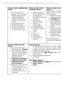
Pulmonary embolism pathophysiology 04.01.01 o Is an obstruction of the pulmonary artery or one of its branches by a thrombus (or thrombi that originates somewhere in the venous system or in the right side of the heart. o Most commonly due to a blood clot or thrombus. o VTE refers to both DVT & PE o Can also be caused by fat. Air, amniotic fluid and septic. Pulmonary embolism clinical manifestations 04.01.01 o Symptoms depends on the size of the embolus, area of pulmonary artery occluded and type of embolus. o Dyspnea (most frequent symptoms) o Chest pain (usually sudden) o Syncope (fainting) o Hemoptysis (coughing up blood) o anxiety o fever o tachycardia o apprehension o cough, diaphoresis (sweating) Pulmonary embolism treatmen 04.01.02 treatment goal is to dissolve (lys existing emboli and prevent new from forming. o Depends on whether pt i hemodynamically stable. o Nasal Oxygen (if pt is sta o Hemodynamically unstab requires emergency trea (hypotension, tachycardi confusion, cardiovascula collapse. o Emergency measures ini improve respiratory & cardiovascular status o Thrombolytic therapy to emboli and return hemodynamic function o Surgical embolectomy & transve catheter embolectomy. Pulmonary embolism prevention 04.01.03 o Pts at risk for PE, the most effective approach is prevention o Active leg exercises to avoid venous stasis, early ambulation and the use of anti-embolism stockings are general preventive measures o Instructs pt to move the legs in a “pumping” exercise so that the leg muscles can help increase venous flow o Advise pt to not sit or lie in bed for prolonged periods, not cross the legs and not to wear constrictive clothing o foot should rest on floor and not dangled. Nursing management o During thrombolytic infusion pt muse remain on bedrest, vit must be assessed frequently, invasive procedures avoided. L ( INR and aPTT must be checked (3-4 hours after) to confirm activation of fibronylic. o Manage chest pain, oxygen therapy, anxiety, assess for signs hypoxemia. o Monitor for potential complications. o Semi- fowlers. Pharmacology Management o Alteplase, Reteplase, Tenecplase (thrombolytic therapy) o Heparin - lovenox o warfarin ARDS clinical manifestations 04.02.01 Acute respiratory distress syndrome. o Rapid onset of severe dyspnea that occurs less than 72 hours after the precipitating event o Arterial hypoxemia that does not respond to supplemental oxygen o Pulmonary edema & bilateral infiltrates seen on chest x-ray. o Acute lung injury then progresses to fibrosing alveolitis with persistent, severe hypoxemia o Increased alveolar dead space o Decreased pulmonary compliance (stiff lungs) ARDS mechanical ventilation 04.02.02 o Primary focus includes identification & treatment of the underlying condition. o Intubation, mechanical ventilation with PEEP to keep alveoli open. o Treat hypovolemia to keep hemodynamically stable. o Prone positioning for best oxygen, frequent repositioning to safeguard integumentary. o Nutritional support, external feedings preferers. o Reduce anxiety, sedation and paralysis o Questions: which is a sign of ARDS? o Heart rate of 140 o Nausea and vomiting o Bloody sputum o Temperature of 98 degree ARDS nursing care of patients on ventilators 04.02.02 o PEEP causes increase end-expiratory pressure is an unnatural pattern breathing and feels strange to the patient. o Patients may be anxious and fight the ventilator. o Frequent assessment of the pt’s status is necessary to evaluate the effectiveness of treatment o Reducing anxiety is important o In some cases, sedation may be required to decrease the patient’s oxygen consumption, allow the ventilator to provide full support of ventilation and decrease the patient’s anxiety o Sedatives that may be used (lorazepam, midazolam(versed), dexmedetomidine (precedex), profodol (diprivan). Mechanical ventilator medication Sedatives that may be used o Lorazepam (Ativan) o Midazolam (versed) o Dexmadetomidien (precede) o Propofol (diprivan) If PEEP Level cannot be maintained despite the use of sedatives neuromuscular blocking agents may be given to paralyze the patient o Pancuronium (pavulon) vecuronium (norcuron) atracurium (tracium) Pulmonary edema/failure nursing interventions 04.03.01 o Positioning the patient to promote circulation ▪ Positioned upright, preferably with the legs dangling over the side of the bed. o Providing psychological support o As the ability to breathe decreases, the patient’s fear and anxiety rise proportionately, making the condition more sever ▪ Reassuring the patient and providing skillful anticipatory nursing care and integral ▪ Patient is is an unstable condition, the nurse remain with the patient. ▪ Nurse gives the patient simple, concise information in a reassuring voice about with is being done to treat the condition and the expected results. o Monitoring medications ▪ Patient receiving diuretic therapy may excrete large volume of urine within minutes after a potent diuretic is given. ▪ Bedside commode may be used to decrease the energy required by the patient ▪ Reduce the resultant increase in cardiac workload induced by getting on and off a bedpan ▪ If necessary, in order to carefully monitor urine output and indwelling urinary catheter may be inserted ▪ Continuous ECG monitoring and frequent measure of vital signs ▪ Patients who received continuing therapy require management in an ICU. ▪ Hypoxemia may occur Pulmonary edema/ failure treatments o Clinical management of a patient with acute pulmonary edema due to left ventricular failure is directed toward reducing volume overload, improving ventricular function and increasing oxygenation. ▪ Goals are accomplished through a combination of oxygen and ventilatory support, iv medication and nursing assessment and interventions o Oxygen therapy o Given in concentrations adequate to relieve hypoxemia and dyspnea o Nonrebreathing mask is used initially o If respiratory failure is severe or persists, noninvasive positive ventilations is the preferred mode of assisted ventilation o Diuretics o Furosemide o Blood pressure is monitored as uring output increase, as it possible for the patient to become hypotensive as intravascular volume decreases o Intake and output, daily weights, serum electrolytes and creatinine are carefully monitored o Vasodilators o Iv nitroglycerin or nitroprusside may enhance symptom relief in pulmonary edema. o Use in contraindicated in patients who are hypotensive o Blood pressure is continually assessed in patients receiving iv Compensated = PH is normal lungs regulate CO2 and thus carbonic acid Bicarbonate-carbonic acid buffer system. Kidneys regulate bicarbonate (HCO3) in ECF Metabolic acidosis (treatments) Causes of Metabolic acidosis -treatment is directed at correcting the Chronic metabolic imbalance chronic renal failure - bicarbonate is given Low calcium is usually with chronic state. - hyperkalemia may occur with acidosis and Acute hypokalemia with reversal of the acidosis Diarrhea with movement of potassium back into the Renal failure – (+acid) cells In kidney failure they are unable to do this resulting in - low serum calcium’s levels are treated excess acid before the chronic metabolic acidosis to Diabetes, lower intestinal fistulas, ureterostomies avoid tetany resulting from an increase in PH and a decrease in ionized calcium - monitor sodium levels - when pH flipped it can make calcium lower. Hypocalcemia may occur with metabolic acidosis Type 1 diabetes- no insulin production The use of diuretics (excessive administration of chloride and parental nutrition without bicarbonate- producing solute. Clinical manifestions o Headache o Confusion/drowsiness o Nausea/vomiting o Severe hypotension (bloop pressure) o Cold, clammy skin o Dysthymias o Hyperkalemia (due to kidney failure) o Chronic metabolic acidosis is usually seen with chronice kidney disease. Balance electrolyte imabalances with ABG Metabolic acidosis o Eliminate the source of the chloride o Sodium bicarbonate during cardiac arrest can result in paradoxical intracellular acidosis o Calcium FIRST (when PH flips it can make calcium lower o Sodium bicarbonate (usually iv) o Monitor sodium levels o Monitor for hypernatremia, hypokalemia, hypocalcemia Normal anion gap is 8-12mEq/l If potassium is included, the normal anion gap is 12- 16mEq/l Metabolic Alkalosis (Treatments.) o treatment is aimed at correcting the underlying acid-base disorder o due to volume depletion for GI loss, the patient’s I&O must be monitored carefully o restore normal fluid volume loss by administering sodium chloride fluids (from puking, give iV) o Patients with HYPOKALEMIA, POTASSIUM as KCI to replace K+ and CI- losses. o H2 receptor antagonists, such as cimetidine (Tagamet), reduce the production of gastric hydrogen chloride (HCI), decreasing the metabolic associated with gastric suction. Causes of Metabolic Alkalosis o Vomiting or gastric suction with loss of hydrogen and chloride ion o Occurs in pyloric stenosis, in which only gastric fluid is lost. o Hypokalemia o Upper Gi secretion loss o Upper Gi secretions are acidic- vomiting or NG tube suctions can result in loss of acids o Antacid overuse o Excess intake of bicarbonate o Hypokalemia will produce alkalosis Chronic cause o Long term diuretics use o Clinical manifestations o Is manifested by symptoms related to decrease calcium ionization o Tingling of the fingers and toe (hypocalcemia) o Dizziness o Hypertonic muscles o Dysrhythmias (hypokalemia) o Atrial tachycardia (increased RR) Management o Restoring normal fluid volume by administering sodium chloride fluids o Hypokalemia, potassium is given as KCI to replace both K+ and CI- losses o H2 receptor antagonists, such as cimetidine (Tagamet) o Monitor I & O carefully o Restore normal fluid volume loss (from puking IV) o Potassium chloride (KCI) (also have chloride losses from vomiting) Hepatitis mode of transmission A & E = Fecal – oral route B & C= bloodborne D = only people with hep B are at risk o Hepatiits A ▪ Ingestion of food or liquids infected with the virus ▪ Spread by poor hand hygiene, usually more likely to be found in daycare settings o Prevention ▪ Good hand hygiene, safe water and proper sewage disposal. o Hepatitis B o Can be found in blood, saliva, semen and vaginal secretions o Transferred from mother to infant (at birth due to close contact) o Tattooing, IV/injection drug use, multiple sexual partners, hemodialysis. o Hepatitis C o Blood transfusions, sexual contact, IV drug use, healthcare needlestick injuries o Hepatitis D o Parenteral spread, IV drug users, hemodialysis and multiple blood transfusions. Hepatitis medication induced ACETAMINOPHEN Hepatitis nutrition o Hepatitis A and B dietary ▪ Advise patient to avoid substances (medications, herbs, illicit drugs and toxins) that may affect liver function such as st.John wort in patients taking hepatitis C virus protases inhibitors ▪ Be aware the enteral feedings may be necessary if anorexia, nausea and vomiting persist. ▪ Carefully monitor fluid balance ▪ Instruct patient to abstain from alcohol during acute illness and for at least 6 months after recovery ▪ Recommended small, frequent meals. Pancreatitis pathophysiology o Inflammation of the pancreas Acute pancreatitis o Self-digestion of the pancreas by its own proteolytic enzymes, principally trypsin, causes acute pancreatitis. Acute pancreatitis clinical manifestations. o Severe abdominal pain is the major symptoms that causes the patient to seek medical care. o Abdominal pain, tenderness and back pain result from irritation and edema of the inflamed pancreas o Pain occurs in the mi epigastrium o Nausea and vomiting o hypotension Pancreatitis treatment o Relieving pain and discomfort o No oral intake including meds o Cimetidine (drug that can be used ) o Proton-pump inhibitors for patients who can’t tolerate h2- antagonist o Parental opioids such as morphine, hydromorphone or fentanyl o Improving breathing pattern o Improving nutritional status o Maintaining skin integrity Pancreatis complications o Fluid and electrolyte are common complications due to nausea, vomiting, movement of fluid from the vascular compartment to the peritoneal cavity o Pancreatic necrosis o Septic shock. o Nurse assesses the patient’s fluid by noting skin turgor and moistness of mucous membranes o Weight the patient daily o Assess the patient for ascites and measures abdominal firth daily o Low serum calcium and magnesium levels may occur and require prompt treatment Chronic pancreatitis causes o Alcoholism and malnutrition are the major causes of chronic pancreatitis o Excessive prolong consumption of alcohol accounts for approximately 70-80% of all cases o Smoking is another cause of chronic pancreatitis’ cause. Chronic pancreatitis manifestation o Characterized by recurring attacks of abdominal pain, and vomiting o Progression of the diseases results in more severe and frequent attacks of pain, longer in duration o Significant weight loss caused by decreased dietary intake secondary to anorexia or fear of eating o Malabsorption occurs late in the disease o Frothy and foul-smelling stool because of impaired fat digestion- steatorrhea o Calcification of the gland and calcium stones may form within the ducts Chronic pancreatitis treatment o Medical Management o Treatment is directed towards: o preventing and managing acute attack o relieving pain and discomfort o Managing exocrine and endocrine insufficiency o Endoscopy o Management of abdominal pain o use of nonopioid measures in early stages but opiods may be used is pain is uncontrolled o Surgical management Cirrhosis Pathophysiology o Is chronic disease characterized by replacement of normal liver tissue with diffuse fibrosis that disrupts the structure and function of the liver. 3 types o Alcoholic cirrhosis ▪ scar tissue characteristically surrounds the portal areas; most frequently caused by chronic alcoholism o postnecrotic cirrhosis ▪ acute viral hepatitis cirrhosis treatment o Many patients with ESLD and cirrhosis use herb milk thistle to treat jaundice and other symptoms; herb has healing and regenerative properties for liver disease o High calorie- sodium restricted Cirrhosis clinical treatment o Promoting rest o Reducing risk of injury o Monitoring and managing potential complications (bleeding and hemorrhage, fluid volume excess) Acute Renal failure classification o 5-point system o RIFLE stands for RISK, INJURY, FAILURE, LOSS AND ESKD (end of stage kidney disease) o RISK, INJURY and FAILURE are considered grades of AKI severity o AKI and SEVERITY are considered outcome of loss. Acute renal failure. PRE/INTRA/POST RENAL Categories of diminished blood flow to the kidneys o PRERENAL o HEMORRHAGE/BLOOD LOSS (diminished blood flow to the kidneys (hypoperfusion). o INTRARENAL o ACTUAL DAMAGE TO KIDNEY DISEASE ▪ Nephrotoxic, diabetes and hypertension, heart failure o POSTRENAL o OBSTRUCTION TO URINE FLOW ▪ Benign prostatic hyperplasia, blood clots, calcite (stones), strictures and tumors. Acute renal failure precautions o Prevention o Increased age o Comorbid conditions o Pre-existing kidney and vascular diseases o Prevention of AKI is essential o Careful history to identify exposure to nephrotoxic agents or environmental toxins o Kidneys are susceptible to the adverse effects of medications as the metabolic by products of most medications are excreted by the kidneys o Patients taking nephrotoxic medications ▪ Aminoglycosides ▪ Gentamicin (garamycin) ▪ Tobramycin ▪ Colistemethae (coly-mycin) ▪ Polymyxin B ▪ Amphotericin B ▪ Vancomycin ▪ Amikacin ▪ Cyclosporine ▪ Should be monitored closely for changes in renal function o Increased age, preexisting kidney disease and the simultaneous administration of several nephrotoxic agents increase the risk of kidney damage o patient WITH HEART FAILURE OR CIRRHOSIS are at particular risk for NSAID induced kidney disease. • As GFR decreases, the creatinine clearance decreases and Serum creatinine and BUN increases • Sodium and water retention • Pts may elect not to undergo dialysis or transplantation. Conservative management and palliative care/ supportive care may be considered • Measures include nutritional therapy, fluid control, medications such as phosphate binders Hemodialysis • Cleansed blood is returned to the body with the goal of removing fluid, balancing electrolytes, and managing acidosis • The dialysate is warmed to body temp to prevent patient discomfort and abdominal pain and to dilate the vessels of the peritoneum to increase urea clearance • Dry heating (heating cabinet, incubator, or heating pads) recommended • Patients on dialysis need higher intake of protein than normal adults High-biologic value proteins Renal failure pharmacology management o Hyperkalemia is the most life-threatening of the fluid and electrolyte changes that occurs in pts with kidney disorders o Administer cation exchange resins such as sodium polystyrene PO • Regulation of protein intake, fluid intake to balance fluid losses, and sodium and potassium restriction Hemodialysis Access • Arteriovenous Fistula (AV) • Preferred access • Arteriovenous graft • Tunneled catheter
Información del documento
- Subido en
- 27 de abril de 2023
- Número de páginas
- 13
- Escrito en
- 2022/2023
- Tipo
- Examen
- Contiene
- Preguntas y respuestas