CELL STRUCTURE
(3.1-3.8)
3.1 Methods of studying cells
Microscopy
What are microscopes? Instruments that produce a magnified image of an
object.
Microscope lenses work more effectively if the convex glass lenses are used in
pairs in a compound light microscope.
● Light microscopes - the relatively long wavelength of light rays means
that a light microscope can only distinguish between two objects if they
are 0.2μm, or further apart.
This limitation can be overcome by using beams of ELECTRONS rather than
beams of light.
● Electron microscopes - with their shorter wavelengths, a beam of
electrons in the electron microscope can distinguish between two
objects only 0.1nm apart.
Magnification
The material that is put under a microscope is referred to as the object. The
appearance of this material when viewed under the microscope is referred to
as the image.
,Magnification: the magnification of an object is how many times bigger the
image is when compared to the size of the real object.
Magnification = size of image/size of real object
Size of real object = size of image/magnification
When calculating magnification, make sure that the units of length are the
same for both the object and the image!
Resolution
Resolution/resolving power: the resolution or resolving power of a microscope
is the minimum distance apart that two objects can be in order for them to
appear as separate items.
● The resolving power depends on the wavelength or form of radiation
used.
● A light microscope has a resolution of about 0.2μm (any two objects that
are 0.2μm or more apart will be seen separately but if they are closer
than this, they will appear as a single item).
● Greater resolution means greater CLARITY.
● The image produced with a high resolution is clearer and more PRECISE.
● Increasing magnification increases the size of the image but doesn’t
always increase the resolution.
● Every microscope has a limit of resolution. Up to this point, increasing
the magnification will reveal more detail, but beyond this point
increasing the magnification will not do this.
● The object will still appear larger but more blurred instead!
, Cell fractionation
To study the structure and function of the various organelles that make up
cells, it is necessary to obtain large numbers of ISOLATED organelles.
Cell fractionation: the process where cells are broken up and the different
organelles they contain are separated out.
Process of cell fractionation:
1. Homogenation
2. Ultracentrifugation
BEFORE cell fractionation begins, the tissue is placed in a cold, buffered
solution which has the same water potential as the tissue. The solution is:
● Cold - to reduce enzyme activity that might break down the organelles.
● Buffered - so that the pH does not fluctuate. Any change in pH could
affect the functioning of enzymes and also alter the structure of the
organelles.
● Of the same water potential as the tissue - to prevent the organelles
from shrinking or bursting due to osmotic loss or gain of water.
Stages of cell fractionation:
1. HOMOGENATION
- The cells are broken up inside a homogeniser (blender)
, - The various organelles from the cell are released.
- The resulting fluid is called the homogenate.
- The homogenate is filtered to remove any complete cells and large
pieces of debris.
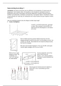
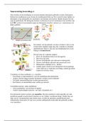
2. ULTRACENTRIFUGATION
Ultracentrifugation: the process by which the fragments in the filtered
homogenate are separated in a machine called a centrifuge.
The centrifuge spins tubes of homogenate at very high speeds in order to
create a centrifugal force.
For animal cells, the process goes like this:
○ The tube of filtrate is placed in the centrifuge and spun at a slow speed.
○ The heaviest organelles i.e. the nuclei are forced to the bottom of the
tube where they form a thin sediment or pellet.
○ The fluid at the top, supernatant, is then removed leaving only the
sediment of nuclei.
○ The supernatant is transferred to another tube and spun in the
centrifuge at a FASTER speed than before.
○ The next heaviest organelles i.e. the mitochondria are forced to the
bottom of the tube.
○ The process is continued in this way so that, at each increase in speed,
the next heaviest organelle is sedimented and separated out.
(3.1-3.8)
3.1 Methods of studying cells
Microscopy
What are microscopes? Instruments that produce a magnified image of an
object.
Microscope lenses work more effectively if the convex glass lenses are used in
pairs in a compound light microscope.
● Light microscopes - the relatively long wavelength of light rays means
that a light microscope can only distinguish between two objects if they
are 0.2μm, or further apart.
This limitation can be overcome by using beams of ELECTRONS rather than
beams of light.
● Electron microscopes - with their shorter wavelengths, a beam of
electrons in the electron microscope can distinguish between two
objects only 0.1nm apart.
Magnification
The material that is put under a microscope is referred to as the object. The
appearance of this material when viewed under the microscope is referred to
as the image.
,Magnification: the magnification of an object is how many times bigger the
image is when compared to the size of the real object.
Magnification = size of image/size of real object
Size of real object = size of image/magnification
When calculating magnification, make sure that the units of length are the
same for both the object and the image!
Resolution
Resolution/resolving power: the resolution or resolving power of a microscope
is the minimum distance apart that two objects can be in order for them to
appear as separate items.
● The resolving power depends on the wavelength or form of radiation
used.
● A light microscope has a resolution of about 0.2μm (any two objects that
are 0.2μm or more apart will be seen separately but if they are closer
than this, they will appear as a single item).
● Greater resolution means greater CLARITY.
● The image produced with a high resolution is clearer and more PRECISE.
● Increasing magnification increases the size of the image but doesn’t
always increase the resolution.
● Every microscope has a limit of resolution. Up to this point, increasing
the magnification will reveal more detail, but beyond this point
increasing the magnification will not do this.
● The object will still appear larger but more blurred instead!
, Cell fractionation
To study the structure and function of the various organelles that make up
cells, it is necessary to obtain large numbers of ISOLATED organelles.
Cell fractionation: the process where cells are broken up and the different
organelles they contain are separated out.
Process of cell fractionation:
1. Homogenation
2. Ultracentrifugation
BEFORE cell fractionation begins, the tissue is placed in a cold, buffered
solution which has the same water potential as the tissue. The solution is:
● Cold - to reduce enzyme activity that might break down the organelles.
● Buffered - so that the pH does not fluctuate. Any change in pH could
affect the functioning of enzymes and also alter the structure of the
organelles.
● Of the same water potential as the tissue - to prevent the organelles
from shrinking or bursting due to osmotic loss or gain of water.
Stages of cell fractionation:
1. HOMOGENATION
- The cells are broken up inside a homogeniser (blender)
, - The various organelles from the cell are released.
- The resulting fluid is called the homogenate.
- The homogenate is filtered to remove any complete cells and large
pieces of debris.
2. ULTRACENTRIFUGATION
Ultracentrifugation: the process by which the fragments in the filtered
homogenate are separated in a machine called a centrifuge.
The centrifuge spins tubes of homogenate at very high speeds in order to
create a centrifugal force.
For animal cells, the process goes like this:
○ The tube of filtrate is placed in the centrifuge and spun at a slow speed.
○ The heaviest organelles i.e. the nuclei are forced to the bottom of the
tube where they form a thin sediment or pellet.
○ The fluid at the top, supernatant, is then removed leaving only the
sediment of nuclei.
○ The supernatant is transferred to another tube and spun in the
centrifuge at a FASTER speed than before.
○ The next heaviest organelles i.e. the mitochondria are forced to the
bottom of the tube.
○ The process is continued in this way so that, at each increase in speed,
the next heaviest organelle is sedimented and separated out.