Comparative Anatomy and Physiology in Animals
Lecture 4 Blood Flow 21/10/20
Heart as a pump
- Heart creates circulation by causing a pressure difference.
- A periodic muscle contraction increases blood pressure inside the heard and forces blood
out of the lumen.
- Contraction phase= systole, relaxation phase= diastole.
- Blood is forced into specialised circulatory system.
- Some systems seem inefficient or maladapted but function within the constraints of an
animal’s lifestyle.
- Atrial and ventricular systole cause: increase in the blood pressure in the atrium lumen,
reduction in lumen volume causing blood to flow out under pressure into the ventricle, and
back flow is prevented by valves in the veins delivering blood or in the heart.
- Rhythmic depolarisation is caused by a rhythmic depolarisation of the cell membranes of the
constituent muscle cells- this causes muscle contraction. This process is initiated by the
heart’s pacemaker.
- Either myogenic (muscles) or neurogenic (nerves).
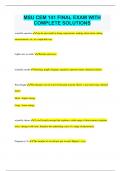
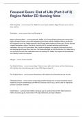
A neurogenic heart (crustaceans and arachnids)
- Neurogenic heart rhythmic depolarisation originates in neurones.
- Each muscle in the heart is innervated and only contracts when stimulated by nerve
impulses.
- The heart has cardiac ganglion- nine neuronal processes have direct nervous contact with
each muscle cell.
- One posterior neuron assumes role of pacemaker- spontaneous periodic train of action
potential that stimulates other neurones, and in turn muscle cells.
Myogenic heart (vertebrates)
- Rhythmic depolarisation originates in muscles.
- Adjacent muscle cells are electrically coupled so depolarisation of one cell rapidly leads to
the direct depolarisation of adjacent cells.
- Cells will spontaneously contract, but not in unison.
- Requires a pacemaker.
- Fish, amphibians, and non-avian reptiles- located in sinus venosus or at its junction with the
atrium.
- Birds and mammals- located in the wall of right atrium.
- Modified muscle cells with reduced contractile apparatus but first to spontaneously
depolarise.
- Depolarisation spreads across myocardium
via conduction.
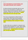
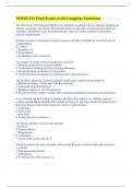
- Mammalian heart - myogenic tissue in
atria are separated from myogenic tissues
in the ventricles by fibrous tissues.
- Connected via the atrioventricular bundle,
which connects with Purkinje fibres.
- Depolarisation of the S-A node leads to a
cascade effect that depolarises the A-V
node.
- Atria contract before the ventricles.
Lecture 4 Blood Flow 21/10/20
Heart as a pump
- Heart creates circulation by causing a pressure difference.
- A periodic muscle contraction increases blood pressure inside the heard and forces blood
out of the lumen.
- Contraction phase= systole, relaxation phase= diastole.
- Blood is forced into specialised circulatory system.
- Some systems seem inefficient or maladapted but function within the constraints of an
animal’s lifestyle.
- Atrial and ventricular systole cause: increase in the blood pressure in the atrium lumen,
reduction in lumen volume causing blood to flow out under pressure into the ventricle, and
back flow is prevented by valves in the veins delivering blood or in the heart.
- Rhythmic depolarisation is caused by a rhythmic depolarisation of the cell membranes of the
constituent muscle cells- this causes muscle contraction. This process is initiated by the
heart’s pacemaker.
- Either myogenic (muscles) or neurogenic (nerves).
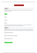
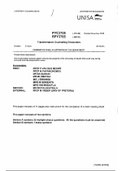
A neurogenic heart (crustaceans and arachnids)
- Neurogenic heart rhythmic depolarisation originates in neurones.
- Each muscle in the heart is innervated and only contracts when stimulated by nerve
impulses.
- The heart has cardiac ganglion- nine neuronal processes have direct nervous contact with
each muscle cell.
- One posterior neuron assumes role of pacemaker- spontaneous periodic train of action
potential that stimulates other neurones, and in turn muscle cells.
Myogenic heart (vertebrates)
- Rhythmic depolarisation originates in muscles.
- Adjacent muscle cells are electrically coupled so depolarisation of one cell rapidly leads to
the direct depolarisation of adjacent cells.
- Cells will spontaneously contract, but not in unison.
- Requires a pacemaker.
- Fish, amphibians, and non-avian reptiles- located in sinus venosus or at its junction with the
atrium.
- Birds and mammals- located in the wall of right atrium.
- Modified muscle cells with reduced contractile apparatus but first to spontaneously
depolarise.
- Depolarisation spreads across myocardium
via conduction.
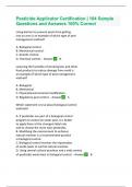
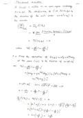
- Mammalian heart - myogenic tissue in
atria are separated from myogenic tissues
in the ventricles by fibrous tissues.
- Connected via the atrioventricular bundle,
which connects with Purkinje fibres.
- Depolarisation of the S-A node leads to a
cascade effect that depolarises the A-V
node.
- Atria contract before the ventricles.