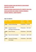
PEDIATRIC ARDMS EXAM AND PRACTICE EXAM NEWEST
2026/2027 TEST BANK
PEDIATRIC ARDMS EXAM REVIEW WITH COMPLETE 200+ REAL
EXAM QUESTIONS AND CORRECT VERIFIED ANSWERS |
ALREADY GRADED A+ (MOST RECENT!)
TABLE OF CONTENTS
Section Domain Approx. Page
Questions Reference
Section Anatomy & Physiology 52 Questions Q1–Q52
I
Section Congenital Variants, 90 Questions Q53–Q142
II Pathology & Pathophysiology
Section Data & Protocols 38 Questions Q143–Q180
III
Section Physics & Instrumentation 10 Questions Q181–Q190
IV
Section Treatment & Emerging 10 Questions Q191–Q200
V Technologies
,SECTION I: ANATOMY AND PHYSIOLOGY
Question 1. A sonographer is performing a neonatal head ultrasound on
a 32-week premature infant. Which of the following structures is BEST
visualized through the anterior fontanelle?
A. Posterior fossa structures
B. The entire ventricular system
C. The spinal cord
D. The optic nerve
CORRECT ANSWER: B
Rationale: The anterior fontanelle serves as the primary acoustic
window for neonatal neurosonography, allowing comprehensive
evaluation of the neonatal brain, including the entire ventricular
system, germinal matrix, and intracranial structures. While the posterior
fontanelle can provide supplementary views of the posterior fossa, the
anterior fontanelle remains the optimal window for visualizing the
ventricular system. The spinal cord and optic nerve require separate
acoustic windows and transducers.
Question 2. Which cranial bone has a characteristic "batwing" shape on
neonatal cranial ultrasound?
A. Frontal bone
B. Parietal bone
C. Sphenoid bone
D. Occipital bone
CORRECT ANSWER: C
,Rationale: The sphenoid bone is characteristically described as having a
"batwing" shape on neonatal cranial ultrasound. This knowledge is
essential for identifying normal cranial anatomy and distinguishing it
from pathological findings. The frontal, parietal, and occipital bones
have different characteristic shapes and positions within the neonatal
skull.
Question 3. A 1-day-old neonate is being evaluated for hydrocephalus.
The anterior fontanelle is used as the acoustic window. Which
anatomical structure is the sonographer primarily evaluating through
this approach?
A. The neonatal spine
B. The neonatal brain
C. The neonatal hip joint
D. The neonatal kidney
CORRECT ANSWER: B
Rationale: The anterior fontanelle serves as the primary acoustic
window for neonatal neurosonography, allowing evaluation of the
neonatal brain. This approach permits visualization of intracranial
structures including the ventricles, germinal matrix, and brain
parenchyma. The neonatal spine, hip joint, and kidney are evaluated
through different acoustic windows and transducer positions.
Question 4. Which of the following is the most common location of
germinal matrix hemorrhage in a preterm infant?
A. Caudothalamic groove
B. Posterior fossa
, C. Subarachnoid space
D. Periventricular white matter
CORRECT ANSWER: A
Rationale: The germinal matrix is located in the caudothalamic groove,
which is the most common site for germinal matrix hemorrhage in
preterm infants. This highly vascular region is particularly vulnerable in
premature neonates due to immature autoregulation of cerebral blood
flow. Posterior fossa hemorrhage, subarachnoid hemorrhage, and
periventricular leukomalacia are distinct entities with different locations
and clinical implications.
Question 5. Which of the following best describes the normal
sonographic appearance of the neonatal thymus?
A. Hyperechoic with multiple calcifications
B. Hypoechoic with a thin echogenic capsule
C. Anechoic with posterior acoustic enhancement
D. Heterogeneous with coarse echotexture
CORRECT ANSWER: B
Rationale: The normal neonatal thymus appears hypoechoic with a thin
echogenic capsule on ultrasound. It is a soft tissue structure located in
the anterior mediastinum. It should not be confused with pathological
entities such as calcifications or cystic structures. The thymus is a
lymphoid organ that is prominent in neonates and involutes with age.
Question 6. Which of the following sutures is formed by the articulation
of the two parietal bones?
2026/2027 TEST BANK
PEDIATRIC ARDMS EXAM REVIEW WITH COMPLETE 200+ REAL
EXAM QUESTIONS AND CORRECT VERIFIED ANSWERS |
ALREADY GRADED A+ (MOST RECENT!)
TABLE OF CONTENTS
Section Domain Approx. Page
Questions Reference
Section Anatomy & Physiology 52 Questions Q1–Q52
I
Section Congenital Variants, 90 Questions Q53–Q142
II Pathology & Pathophysiology
Section Data & Protocols 38 Questions Q143–Q180
III
Section Physics & Instrumentation 10 Questions Q181–Q190
IV
Section Treatment & Emerging 10 Questions Q191–Q200
V Technologies
,SECTION I: ANATOMY AND PHYSIOLOGY
Question 1. A sonographer is performing a neonatal head ultrasound on
a 32-week premature infant. Which of the following structures is BEST
visualized through the anterior fontanelle?
A. Posterior fossa structures
B. The entire ventricular system
C. The spinal cord
D. The optic nerve
CORRECT ANSWER: B
Rationale: The anterior fontanelle serves as the primary acoustic
window for neonatal neurosonography, allowing comprehensive
evaluation of the neonatal brain, including the entire ventricular
system, germinal matrix, and intracranial structures. While the posterior
fontanelle can provide supplementary views of the posterior fossa, the
anterior fontanelle remains the optimal window for visualizing the
ventricular system. The spinal cord and optic nerve require separate
acoustic windows and transducers.
Question 2. Which cranial bone has a characteristic "batwing" shape on
neonatal cranial ultrasound?
A. Frontal bone
B. Parietal bone
C. Sphenoid bone
D. Occipital bone
CORRECT ANSWER: C
,Rationale: The sphenoid bone is characteristically described as having a
"batwing" shape on neonatal cranial ultrasound. This knowledge is
essential for identifying normal cranial anatomy and distinguishing it
from pathological findings. The frontal, parietal, and occipital bones
have different characteristic shapes and positions within the neonatal
skull.
Question 3. A 1-day-old neonate is being evaluated for hydrocephalus.
The anterior fontanelle is used as the acoustic window. Which
anatomical structure is the sonographer primarily evaluating through
this approach?
A. The neonatal spine
B. The neonatal brain
C. The neonatal hip joint
D. The neonatal kidney
CORRECT ANSWER: B
Rationale: The anterior fontanelle serves as the primary acoustic
window for neonatal neurosonography, allowing evaluation of the
neonatal brain. This approach permits visualization of intracranial
structures including the ventricles, germinal matrix, and brain
parenchyma. The neonatal spine, hip joint, and kidney are evaluated
through different acoustic windows and transducer positions.
Question 4. Which of the following is the most common location of
germinal matrix hemorrhage in a preterm infant?
A. Caudothalamic groove
B. Posterior fossa
, C. Subarachnoid space
D. Periventricular white matter
CORRECT ANSWER: A
Rationale: The germinal matrix is located in the caudothalamic groove,
which is the most common site for germinal matrix hemorrhage in
preterm infants. This highly vascular region is particularly vulnerable in
premature neonates due to immature autoregulation of cerebral blood
flow. Posterior fossa hemorrhage, subarachnoid hemorrhage, and
periventricular leukomalacia are distinct entities with different locations
and clinical implications.
Question 5. Which of the following best describes the normal
sonographic appearance of the neonatal thymus?
A. Hyperechoic with multiple calcifications
B. Hypoechoic with a thin echogenic capsule
C. Anechoic with posterior acoustic enhancement
D. Heterogeneous with coarse echotexture
CORRECT ANSWER: B
Rationale: The normal neonatal thymus appears hypoechoic with a thin
echogenic capsule on ultrasound. It is a soft tissue structure located in
the anterior mediastinum. It should not be confused with pathological
entities such as calcifications or cystic structures. The thymus is a
lymphoid organ that is prominent in neonates and involutes with age.
Question 6. Which of the following sutures is formed by the articulation
of the two parietal bones?